. 2022 Apr;12(4):2120-2126.
doi: 10.1016/j.apsb.2021.11.007. Epub 2021 Nov 16.

Unraveling the drug distribution in brain enabled by MALDI MS imaging with laser-assisted chemical transfer

Affiliations

Unraveling the drug distribution in brain enabled by MALDI MS imaging with laser-assisted chemical transfer

Shuai Guo et al. Acta Pharm Sin B. 2022 Apr.

Abstract

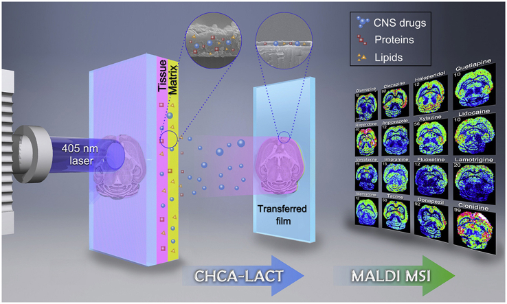
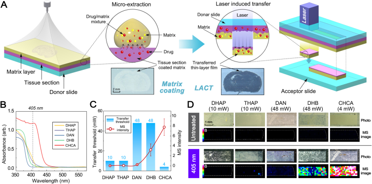
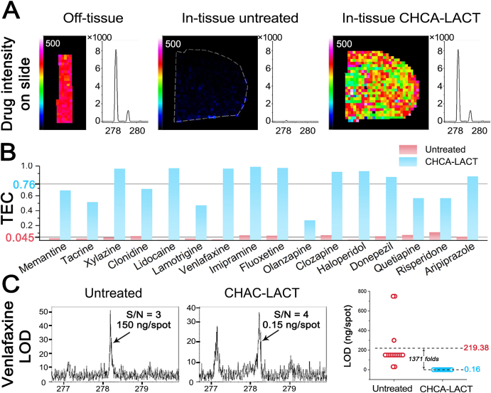
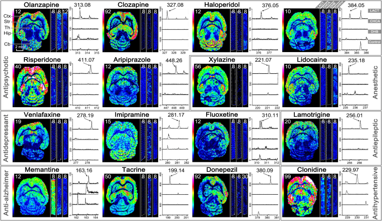
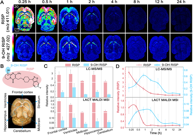
Accurate localization of central nervous system (CNS) drug distribution in the brain is quite challenging to matrix-assisted laser desorption/ionization (MALDI) mass spectrometry imaging (MSI), owing to the ionization competition/suppression of highly abundant endogenous biomolecules and MALDI matrix. Herein, we developed a highly efficient sample preparation technique, laser-assisted chemical transfer (LACT), to enhance the detection sensitivity of CNS drugs in brain tissues. A focused diode laser source transilluminated the tissue slide coated with α-cyano-4-hydroxycinnamic acid, an optimal matrix to highly absorb the laser radiation at 405 nm, and a very thin-layer chemical film mainly containing drug molecule was transferred to the acceptor glass slide. Subsequently, MALDI MSI was performed on the chemical film without additional sample treatment. One major advantage of LACT is to minimize ionization competition/suppression from the tissue itself by removing abundant endogenous lipid and protein components. The superior performance of LACT led to the successful visualization of regional distribution patterns of 16 CNS drugs in the mouse brain. Furthermore, the dynamic spatial changes of risperidone and its metabolite were visualized over a 24-h period. Also, the brain-to-plasma (B/P) ratio could be obtained according to MALDI MSI results, providing an alternative means to assess brain penetration in drug discovery.

Keywords: Brain penetration; Drug distribution; Laser-assisted chemical transfer; Mass spectrometry imaging; Pharmaceutical analysis.


Figures

Image 1
Graphical abstract
Figure 1
Figure 1
Working principle of LACT and optimization of absorbing matrix compound for LACT and MALDI MSI. (A) Workflow for matrix deposition, laser illumination (405 nm), and chemical transfer. (B) Solid-state UV absorption spectra of different MALDI matrices coated on brain tissues. (C) The influence of transfer threshold energy on the signal intensities of venlafaxine. The transfer threshold energy was defined as the minimum real-time power of the laser (mW) necessary to achieve chemical transfer. Error bars indicate the standard deviations of 100 mass spectra acquired from ion images. (D) Comparison of MALDI MSI results of venlafaxine measured in dosed mouse brain before and after LACT treatment using different matrices. DHAP, 2,6-dihydroxyacetophenone; THAP, 2,4,6-trihydroxy-acetophenone; DAN, 1,5-diaminonapthalene; DHB, 2,5-dihydroxybenzoic acid; CHCA, α-cyano-4-hydroxycinnamic acid.
Figure 2
Figure 2
Evaluation of tissue extinction coefficient (TEC) and limit of detection (LOD) of CHCA-LACT technique. (A) Ion images and representative MALDI mass spectra of venlafaxine acquired from bare ITO glass slide, untreated tissue, and chemical film. (B) Histogram of calculated TEC values for each CNS drug in untreated and CHCA-LACT treated brain tissue sections. (C) Representative MALDI mass spectra of venlafaxine at LOD level and LODs of 16 CNS drugs obtained from untreated and CHCA-LACT treated tissue sections.
Figure 3
Figure 3
Comparison of the performance of three MALDI MSI methods from left to right for each CNS drug, including MALDI with CHCA-LACT (whole horizontal section), MALDI with CHCA (vertical stripe), and MALDI with DHB (vertical stripe). The blank tissue treated by CHCA-LACT was used as negative control (vertical stripe). Representative mass spectra corresponding to each imaging experiment were shown from top to bottom: MALDI with CHCA-LACT, MALDI with CHCA, MALDI with DHB, and blank control. The value of the color scale bar represents the absolute intensity divided by 10. Ctx, cerebral cortex; Str, striatum; Th, thalamus; Hip, hippocampus; Cb, cerebellum.
Figure 4
Figure 4
Visualization of spatiotemporal changes of risperidone (RISP) and its metabolite 9-hydroxy-risperidone (9-OH RISP) in brain tissues after i.p. administration using MALDI MSI with CHCA-LACT. (A) Ion images of RISP and 9-OH RISP were acquired at different time points. (B) Chemical structures of RISP and 9-OH RISP, and optical image of the horizontal section of the mouse brain. (C) Comparison of relative intensities of RISP and 9-OH RISP at different brain regions determined by LC‒MS/MS and MALDI MSI with CHCA-LACT, respectively. (D) Comparison of relative intensities of RISP and 9-OH RISP in the lateral ventricles at different time points determined by LC‒MS/MS and MALDI MSI with CHCA-LACT, respectively. The results were expressed as the mean ± SD from three animals.

Similar articles

Cited by

References

    1. Mullard A. New drugs cost US $2.6 billion to develop. Nat Rev Drug Discov. 2014;13:877.
    1. Cummings J.L., Morstorf T., Zhong K. Alzheimer's disease drug-development pipeline: few candidates, frequent failures. Alzheimer's Res Ther. 2014;6:1–7. - PMC - PubMed
    1. Morgan P., Brown D.G., Lennard S., Anderton M.J., Barrett J.C., Eriksson U., et al. Impact of a five-dimensional framework on R&D productivity at AstraZeneca. Nat Rev Drug Discov. 2018;17:167. - PubMed
    1. Di L., Kerns E.H., Carter G.T. Strategies to assess blood–brain barrier penetration. Expet Opin Drug Discov. 2008;3:677–687. - PubMed
    1. Wong A.L.A., Xiang X., Ong P.S., Mitchell E.Q.Y., Syn N., Wee I., et al. A review on liquid chromatography-tandem mass spectrometry methods for rapid quantification of oncology drugs. Pharmaceutics. 2018;10:221. - PMC - PubMed

LinkOut - more resources