

Celastrol enhances transcription factor EB (TFEB)-mediated autophagy and mitigates Tau pathology: Implications for Alzheimer's disease therapy
- PMID: 35847498
- PMCID: PMC9279716
- DOI: 10.1016/j.apsb.2022.01.017
Celastrol enhances transcription factor EB (TFEB)-mediated autophagy and mitigates Tau pathology: Implications for Alzheimer's disease therapy
Abstract
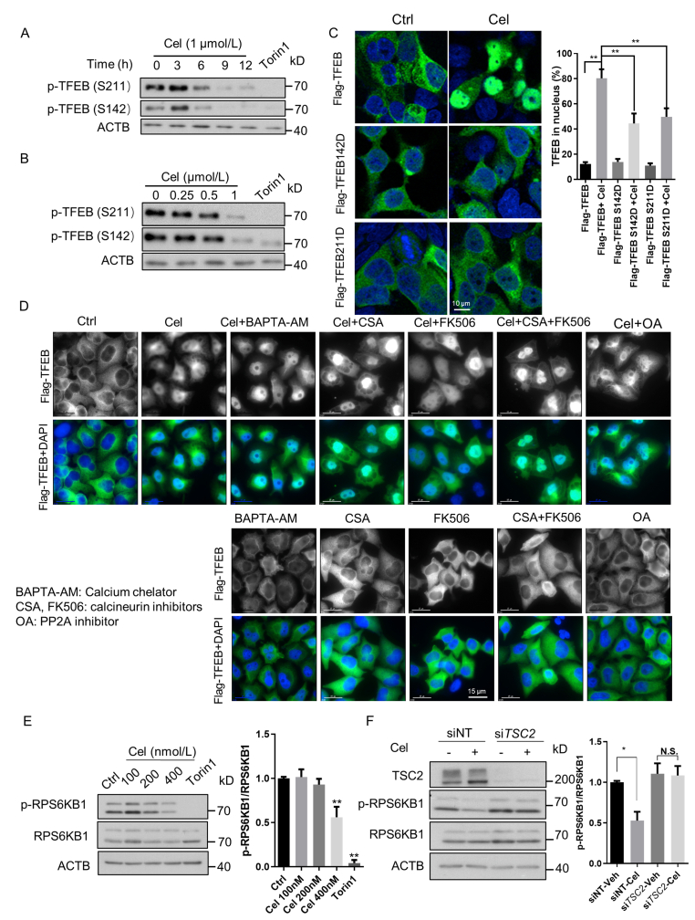
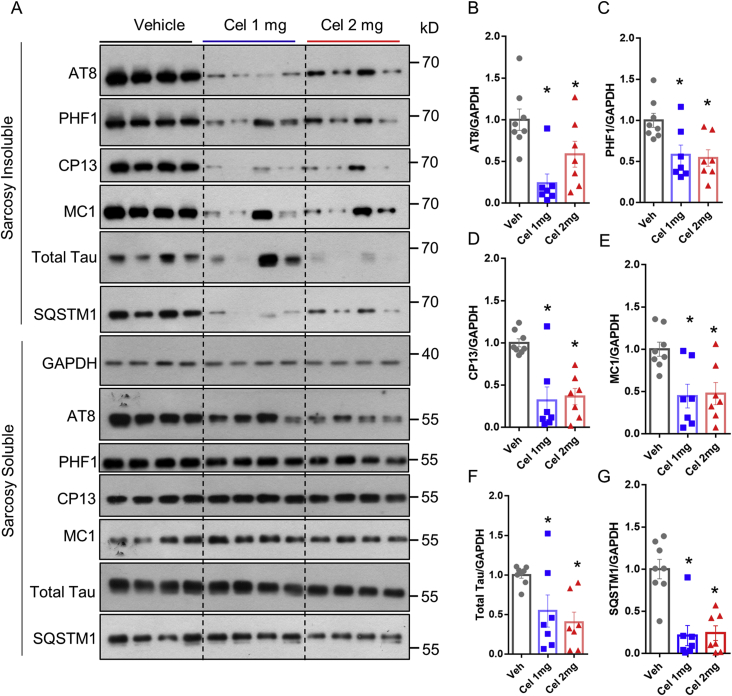
Alzheimer's disease (AD), characterized by the accumulation of protein aggregates including phosphorylated Tau aggregates, is the most common neurodegenerative disorder with limited therapeutic agents. Autophagy plays a critical role in the degradation of phosphorylated Tau aggregates, and transcription factor EB (TFEB) is a master regulator of autophagy and lysosomal biogenesis. Thus, small-molecule autophagy enhancers targeting TFEB hold promise for AD therapy. Here, we found that celastrol, an active ingredient isolated from the root extracts of Tripterygium wilfordii (Lei Gong Teng in Chinese) enhanced TFEB-mediated autophagy and lysosomal biogenesis in vitro and in mouse brains. Importantly, celastrol reduced phosphorylated Tau aggregates and attenuated memory dysfunction and cognitive deficits in P301S Tau and 3xTg mice, two commonly used AD animal models. Mechanistical studies suggest that TFEB-mediated autophagy-lysosomal pathway is responsible for phosphorylated Tau degradation in response to celastrol. Overall, our findings indicate that Celastrol is a novel TFEB activator that promotes the degradation of phosphorylated Tau aggregates and improves memory in AD animal models. Therefore, Celastrol shows potential as a novel agent for the treatment and/or prevention of AD and other tauopathies.
Keywords: Alzheimer's disease (AD); Autophagy; Celastrol; Lysosome biogenesis; TFEB; Tau; Therapeutic target; mTOR.
© 2022 Chinese Pharmaceutical Association and Institute of Materia Medica, Chinese Academy of Medical Sciences. Production and hosting by Elsevier B.V.
Figures


Comment in
-
Celastrol, a TFEB (transcription factor EB) agonist, is a promising drug candidate for Alzheimer disease.Autophagy. 2022 Jul;18(7):1740-1742. doi: 10.1080/15548627.2022.2046437. Epub 2022 Mar 6. Autophagy. 2022. PMID: 35253615 Free PMC article.
Similar articles
-
Celastrol, a TFEB (transcription factor EB) agonist, is a promising drug candidate for Alzheimer disease.Autophagy. 2022 Jul;18(7):1740-1742. doi: 10.1080/15548627.2022.2046437. Epub 2022 Mar 6. Autophagy. 2022. PMID: 35253615 Free PMC article.
-
A small molecule transcription factor EB activator ameliorates beta-amyloid precursor protein and Tau pathology in Alzheimer's disease models.Aging Cell. 2020 Feb;19(2):e13069. doi: 10.1111/acel.13069. Epub 2019 Dec 19. Aging Cell. 2020. PMID: 31858697 Free PMC article.
-
Theranostic F-SLOH mitigates Alzheimer's disease pathology involving TFEB and ameliorates cognitive functions in Alzheimer's disease models.Redox Biol. 2022 May;51:102280. doi: 10.1016/j.redox.2022.102280. Epub 2022 Mar 8. Redox Biol. 2022. PMID: 35286997 Free PMC article.
-
Therapeutic Potential of Vital Transcription Factors in Alzheimer's and Parkinson's Disease With Particular Emphasis on Transcription Factor EB Mediated Autophagy.Front Neurosci. 2021 Dec 14;15:777347. doi: 10.3389/fnins.2021.777347. eCollection 2021. Front Neurosci. 2021. PMID: 34970114 Free PMC article. Review.
-
TFEB in Alzheimer's disease: From molecular mechanisms to therapeutic implications.Neurobiol Dis. 2022 Oct 15;173:105855. doi: 10.1016/j.nbd.2022.105855. Epub 2022 Aug 27. Neurobiol Dis. 2022. PMID: 36031168 Review.
Cited by
-
Transcriptional regulation of autophagy and its implications in human disease.Cell Death Differ. 2023 Jun;30(6):1416-1429. doi: 10.1038/s41418-023-01162-9. Epub 2023 Apr 12. Cell Death Differ. 2023. PMID: 37045910 Free PMC article. Review.
-
Enhancement of lysosome biogenesis as a potential therapeutic approach for neurodegenerative diseases.Neural Regen Res. 2023 Nov;18(11):2370-2376. doi: 10.4103/1673-5374.371346. Neural Regen Res. 2023. PMID: 37282455 Free PMC article.
-
Autophagic mechanisms in longevity intervention: role of natural active compounds.Expert Rev Mol Med. 2023 Mar 30;25:e13. doi: 10.1017/erm.2023.5. Expert Rev Mol Med. 2023. PMID: 36994671 Free PMC article. Review.
-
Pathogenesis of Alzheimer's disease and therapeutic strategies involving traditional Chinese medicine.RSC Med Chem. 2024 Oct 3;15(12):3950-69. doi: 10.1039/d4md00660g. Online ahead of print. RSC Med Chem. 2024. PMID: 39430949 Free PMC article. Review.
-
Drofenine as a Kv2.1 inhibitor alleviated AD-like pathology in mice through Aβ/Kv2.1/microglial NLRP3/neuronal Tau axis.Acta Pharm Sin B. 2025 Jan;15(1):371-391. doi: 10.1016/j.apsb.2024.11.010. Epub 2024 Nov 26. Acta Pharm Sin B. 2025. PMID: 40041886 Free PMC article.
References
-
- 2020 Alzheimer's disease facts and figures. Alzheimers Dement. 2020;16:391–460.
-
- Hardy J. A hundred years of Alzheimer's disease research. Neuron. 2006;52:3–13. - PubMed
-
- Iyaswamy A., Krishnamoorthi S.K., Zhang H., Sreenivasmurthy S.G., Zhu Z., Liu J., et al. Qingyangshen mitigates amyloid-β and Tau aggregate defects involving PPARα-TFEB activation in transgenic mice of Alzheimer's disease. Phytomedicine. 2021;91:153648. - PubMed
LinkOut - more resources
Full Text Sources
Miscellaneous
