Review
. 2022 Jul 5:2022:3345637.
doi: 10.1155/2022/3345637. eCollection 2022.

Biological Effects and Mechanisms of Caspases in Early Brain Injury after Subarachnoid Hemorrhage

Affiliations
Review

Biological Effects and Mechanisms of Caspases in Early Brain Injury after Subarachnoid Hemorrhage

Yiwen Wu et al. Oxid Med Cell Longev. .

Abstract

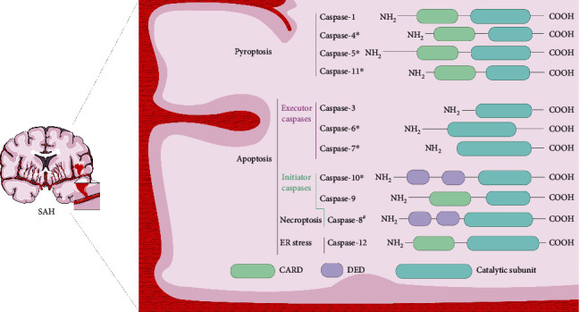
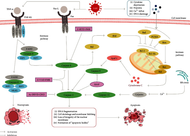
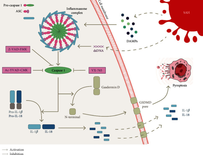
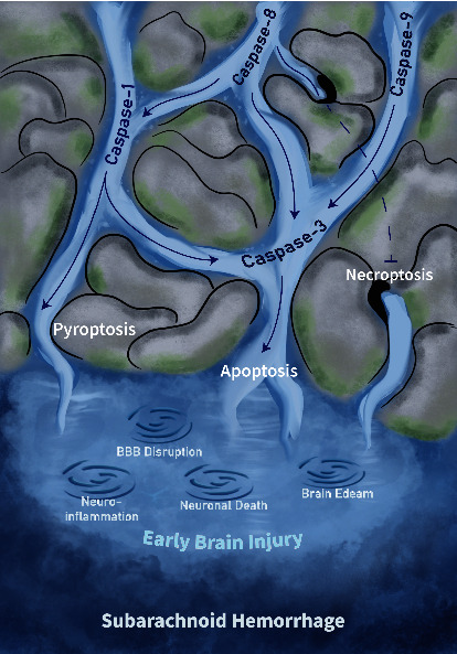
Caspases are an evolutionarily conserved family of proteases responsible for mediating and initiating cell death signals. In the past, the dysregulated activation of caspases was reported to play diverse but equally essential roles in neurodegenerative diseases, such as brain injury and neuroinflammatory diseases. A subarachnoid hemorrhage (SAH) is a traumatic event that is either immediately lethal or induces a high risk of stroke and neurological deficits. Currently, the prognosis of SAH after treatment is not ideal. Early brain injury (EBI) is considered one of the main factors contributing to the poor prognosis of SAH. The mechanisms of EBI are complex and associated with oxidative stress, neuroinflammation, blood-brain barrier disruption, and cell death. Based on mounting evidence, caspases are involved in neuronal apoptosis or death, endothelial cell apoptosis, and increased inflammatory cytokine-induced by apoptosis, pyroptosis, and necroptosis in the initial stages after SAH. Caspases can simultaneously mediate multiple death modes and regulate each other. Caspase inhibitors (including XIAP, VX-765, and Z-VAD-FMK) play an essential role in ameliorating EBI after SAH. In this review, we explore the related pathways mediated by caspases and their reciprocal regulation patterns after SAH. Furthermore, we focus on the extensive crosstalk of caspases as a potential area of research on therapeutic strategies for treating EBI after SAH.


Conflict of interest statement

The authors declare no conflict of interest.

Figures

Figure 1
Figure 1
Functional classification and domain architecture of caspases in SAH. Caspases are classified in SAH as pyroptotic or apoptotic caspases according to their function and domain structure. Apoptotic caspases can be further subdivided into initiator caspases and executioner caspases. The function of initiating caspases is to initiate apoptosis by activating executioner caspases. Furthermore, necroptosis occurs when caspase-8 is inhibited or knocked out. Caspase-12 is structurally like inflammatory caspases but may be activated by endoplasmic reticulum stress to participate in apoptosis. Until today, caspase-4, caspase-5, caspase-6, caspase-7, caspase-10, and caspase-11 have not been experimentally demonstrated in SAH, and their roles in SAH remain undefined. #Inhibition of caspase-8 activates necroptosis.
Figure 2
Figure 2
Apoptosis and necroptosis in SAH. The intrinsic pathway is activated by various stresses after SAH prompting activation of Bak and Bax and release of cytochrome c, which binds to Apaf-1 and then activates through self-cleavage of procaspase-9, further initiating the apoptotic cascade of caspase-3. The extrinsic pathway is activated through membrane receptor formation complexes, and the programmed death cascade induced through caspase-8 to caspase-3 activation. In addition, endoplasmic reticulum stress activates caspase-12 via calcium-activated calpains and ultimately acts on caspase-3 to mediate apoptosis. The natural inhibitor XIAP, the broad-spectrum inhibitor Z-VAD-FMK, the artificial inhibitors Z-IETD-FMK and Ac-DEVD-CHO, which exert inhibitory functions on caspase-8, caspase-9, and caspase-3, respectively, can effectively alleviate apoptosis in EBI. Necroptosis is triggered downstream of the death domain receptor TNFR. Upon detection of a “death signal,” RIP1 is activated and the subsequent formation of a RIP homotypic interaction motif (RHIM) begins to recruit RIP3. The RIPK1/RIP3 complex recruits and phosphorylates MLKL to form necrosomes. Ultimately, MLKL acts on the cell membrane to form pores that lead to necroptosis by allowing the uncontrolled release of intracellular substances.
Figure 3
Figure 3
Pyroptosis pathway in SAH. Pyroptosis is an inflammatory form of cell death that is stimulated in SAH by intracellular sensor proteins such as NLRP3 (used as an example here) or AIM2 that detect damage-associated molecular patterns (DAMPs) or cytoplasmic double-stranded DNA (dsDNA). Upon receiving these stimuli, these sensors recruit an adaptor, the apoptosis-related speck-like protein ASC, which in turn recruit procaspase-1 to form the inflammasome complex and activate caspase-1 within it. The activation of caspase-1 cleaves precursors of the interleukin-1 family cytokines IL-1β and IL-18. In addition, caspase-1 also activates gasdermin D (GSDMD), causing its N-terminal domain to be cleaved and forming a pore (GSDMD pore) in the plasma membrane. Mature IL-1β and IL18 are released along the GSDMD pore and cause cell swelling eventually triggering pyroptosis. Moreover, both the broad-spectrum caspase inhibitor Z-VAD-FMK and caspase-1-specific inhibitors (VX-765 and Ac-YVAD-CMK) can alleviate EBI after SAH by inhibiting the caspase-1-mediated pyroptosis pathway.
Figure 4
Figure 4
Relationship between caspase-mediated programmed cell death's crosstalk and the underlying pathological mechanisms in EBI.

References

    1. Van Opdenbosch N., Lamkanfi M. Caspases in cell death, inflammation, and disease. Immunity . 2019;50(6):1352–1364. doi: 10.1016/j.immuni.2019.05.020. - DOI - PMC - PubMed
    1. Van Gorp H., Van Opdenbosch N., Lamkanfi M. Inflammasome-dependent cytokines at the crossroads of health and autoinflammatory disease. Cold Spring Harbor Perspectives in Biology . 2019;11(1) doi: 10.1101/cshperspect.a028563. - DOI - PMC - PubMed
    1. Burguillos M. A., Deierborg T., Kavanagh E., et al. Caspase signalling controls microglia activation and neurotoxicity. Nature . 2011;472(7343):319–324. doi: 10.1038/nature09788. - DOI - PubMed
    1. Devlin P., Ishrat T., Stanfill A. G. A systematic review of inflammatory cytokine changes following aneurysmal subarachnoid hemorrhage in animal models and humans. Translational Stroke Research . 2022 doi: 10.1007/s12975-022-01001-y. - DOI - PubMed
    1. Grüter B. E., Croci D., Schöpf S., et al. Systematic review and meta-analysis of methodological quality in in vivo animal studies of subarachnoid hemorrhage. Translational Stroke Research . 2020;11(6):1175–1184. doi: 10.1007/s12975-020-00801-4. - DOI - PubMed

LinkOut - more resources