. 2022 Jun 28:9:923356.
doi: 10.3389/fvets.2022.923356. eCollection 2022.

Biomechanical and Microstructural Properties of Subchondral Bone From Three Metacarpophalangeal Joint Sites in Thoroughbred Racehorses

Affiliations

Biomechanical and Microstructural Properties of Subchondral Bone From Three Metacarpophalangeal Joint Sites in Thoroughbred Racehorses

Duncan J Pearce et al. Front Vet Sci. .

Abstract

Fatigue-induced subchondral bone (SCB) injury is common in racehorses. Understanding how subchondral microstructure and microdamage influence mechanical properties is important for developing injury prevention strategies. Mechanical properties of the disto-palmar third metacarpal condyle (MCIII) correlate poorly with microstructure, and it is unknown whether the properties of other sites within the metacarpophalangeal (fetlock) joint are similarly complex. We aimed to investigate the mechanical and structural properties of equine SCB from specimens with minimal evidence of macroscopic disease. Three sites within the metacarpophalangeal joint were examined: the disto-palmar MCIII, disto-dorsal MCIII, and proximal sesamoid bone. Two regions of interest within the SCB were compared, a 2 mm superficial and an underlying 2 mm deep layer. Cartilage-bone specimens underwent micro-computed tomography, then cyclic compression for 100 cycles at 2 Hz. Disto-dorsal MCIII specimens were loaded to 30 MPa (n = 10), while disto-palmar MCIII (n = 10) and proximal sesamoid (n = 10) specimens were loaded to 40 MPa. Digital image correlation determined local strains. Specimens were stained with lead-uranyl acetate for volumetric microdamage quantification. The dorsal MCIII SCB had lower bone volume fraction (BVTV), bone mineral density (BMD), and stiffness compared to the palmar MCIII and sesamoid bone (p < 0.05). Superficial SCB had higher BVTV and lower BMD than deeper SCB (p < 0.05), except at the palmar MCIII site where there was no difference in BVTV between depths (p = 0.419). At all sites, the deep bone was stiffer (p < 0.001), although the superficial to deep gradient was smaller in the dorsal MCIII. Hysteresis (energy loss) was greater superficially in palmar MCIII and sesamoid (p < 0.001), but not dorsal MCIII specimens (p = 0.118). The stiffness increased with cyclic loading in total cartilage-bone specimens (p < 0.001), but not in superficial and deep layers of the bone, whereas hysteresis decreased with the cycle for all sites and layers (p < 0.001). Superficial equine SCB is uniformly less stiff than deeper bone despite non-uniform differences in bone density and damage levels. The more compliant superficial layer has an important role in energy dissipation, but whether this is a specific adaptation or a result of microdamage accumulation is not clear.

Keywords: equine; hysteresis; metacarpus; microdamage; stiffness; subchondral bone.


Conflict of interest statement

The authors declare that the research was conducted in the absence of any commercial or financial relationships that could be construed as a potential conflict of interest.

Figures
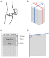
Figure 1
Figure 1
Schematic drawings showing the locations of specimen collection within a fetlock joint (A) and of a cartilage-bone specimen where nonmineralized cartilage is represented in white, and mineralized tissue in gray (B–D). (A) Parasagittal view through the lateral condyle of a fetlock joint, where ‘a’ is the disto-dorsal MCIII, ‘b’ the disto-palmar MCIII, and ‘c’ the proximal sesamoid specimen collection sites. (B) The cylindrical specimen with a flat cut face, and lines ‘A’ and ‘B’. (C) View of the stained face showing the 2 mm thick superficial and deep layers. (D) Sagittal view along line ‘B’ showing the angle used to calculate mineralized tissue surface angle, and the cross-sectional shaded area of hyaline cartilage used to measure surface evenness.
Figure 2
Figure 2
MicroCT images of a palmar MCIII cartilage-bone specimen from a Thoroughbred racehorse with microfractures before (A,C) and after (B,D) staining the bone with lead-uranyl acetate. Image (A) and (B) are short axis images taken approximately 1 mm below the mineralized to non-mineralized cartilage interface. Images (C) and (D) are long axis images of the superficial half of the specimen. Image (A) was taken at an approximately equivalent slice as image (B); likewise, with images (C) and (D). The staining in images (B) and (D) confirms uptake in this specimen with known microdamage (arrows). Resorptive lesions are present (arrowheads).
Figure 3
Figure 3
MicroCT images of a palmar MCIII cartilage-bone specimen from a Thoroughbred racehorse. (A,C) are unlabelled images without evidence of microfractures, while (B,D) are labeled images taken at approximately equivalent slices after staining the bone with lead-uranyl acetate showing small areas of label uptake near the articular surface consistent with microdamage (highlighted by white arrows). Image (A,B) are short axis images taken approximately 0.5 mm below the mineralized to non-mineralized cartilage interface. Images (C,D) are long axis images of the superficial half of the specimen. Resorptive lesions are present (arrowheads).
Figure 4
Figure 4
Boxplots of bone volume fraction (A), bone mineral density (B), damaged bone volume fraction (C), and adjusted damaged bone volume fraction (D) within the metacarpophalangeal joint of Thoroughbred racehorses [n = 10 except for (C) and (D) where n = 9 in the deep dorsal site] stratified by site (palmar MCIII, dorsal MCIII, proximal sesamoid) and by layer (superficial 2 mm, deeper 2 mm, or total ~10 mm thick specimen). BVTV, bone volume fraction; BMD, bone mineral density in mg HA/ccm; DBVF, damaged bone volume fraction; DBV/BSA, adjusted damaged bone volume fraction in mm−2; X represents the mean, the horizontal midpoint of the box the median, the lower end of the box the first quartile, the upper end of the box the third quartile, and the “whiskers” extend from the ends of the box to the maximum and minimum values. The dots signify outlier values. *,** Within each site, layers with different asterisk annotations are significantly different (P < 0.05). Total specimen BVTV and BMD was not compared to superficial and deep as it comprises both. a,b,c Within each layer, sites with different alphabetical annotations are significantly different (P < 0.05).
Figure 5
Figure 5
Stiffness (MPa) of cartilage-bone specimens over the first nine compressive loading cycles at three sites (dorsal MCIII, palmar MCIII and proximal sesamoid) within the metacarpophalangeal joint of n = 10 Thoroughbred racehorses. Layers of each specimen (superficial 2 mm, deeper 2 mm, and total ~10 mm thick cartilage-bone) are stratified into columns. Unadjusted means with 95% confidence intervals are displayed.
Figure 6
Figure 6
Normalized hysteresis (fraction of energy loss) of cartilage-bone specimens over the first nine compressive loading cycles at three sites (dorsal MCIII, palmar MCIII and proximal sesamoid) within the metacarpophalangeal joint of n = 10 Thoroughbred racehorses. Layers of each specimen (superficial 2 mm, deeper 2 mm, and total ~10 mm thick cartilage-bone) are stratified into columns. Unadjusted means with 95% confidence intervals are displayed. Note that in the deep layer, dorsal (blue) hysteresis is obscured by sesamoid (green) hysteresis.

References

    1. Wylie CE, McManus P, McDonald C, Jorgensen S, McGreevy P. Thoroughbred fatality and associated jockey falls and injuries in races in New South Wales and the Australian Capital Territory, Australia: 2009-2014. Vet J. (2017) 227:1–7. 10.1016/j.tvjl.2017.06.008 - DOI - PubMed
    1. Rosanowski SM, Chang YM, Stirk AJ, Verheyen KLP. Epidemiology of race-day distal limb fracture in flat racing thoroughbreds in Great Britain (2000-2013). Equine Vet J. (2019) 51:83–9. 10.1111/evj.12968 - DOI - PubMed
    1. Sun TC, Riggs CM, Cogger N, Wright J, Al-Alawneh JI. Noncatastrophic and catastrophic fractures in racing thoroughbreds at the Hong Kong Jockey Club. Equine Vet J. (2019) 51:77–82. 10.1111/evj.12953 - DOI - PubMed
    1. Spargo KE, Rubio-Martinez LM, Wheeler DP, Fletcher L, Carstens A. Catastrophic musculoskeletal injuries in thoroughbred racehorses on racetracks in Gauteng, South Africa. J S Afr Vet Assoc. (2019) 90:5. 10.4102/jsava.v90i0.1640 - DOI - PMC - PubMed
    1. Parkin IDH, Clegg PD, French NP, Proudman CJ, Riggs CM, Singer ER, et al. . Risk of fatal distal limb fractures among thoroughbreds involved in the five types of racing in the United Kingdom. Vet Rec. (2004) 154:493–7. 10.1136/vr.154.16.493 - DOI - PubMed

LinkOut - more resources