. 2020 May 20;1(1):23-34.
doi: 10.1002/jha2.11. eCollection 2020 Jul.

A pilot clinical phase II trial MemSID: Acute and durable changes of red blood cells of sickle cell disease patients on memantine treatment

Affiliations

A pilot clinical phase II trial MemSID: Acute and durable changes of red blood cells of sickle cell disease patients on memantine treatment

Asya Mahkro et al. EJHaem. .

Abstract

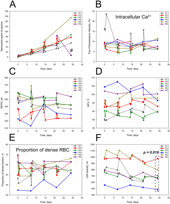
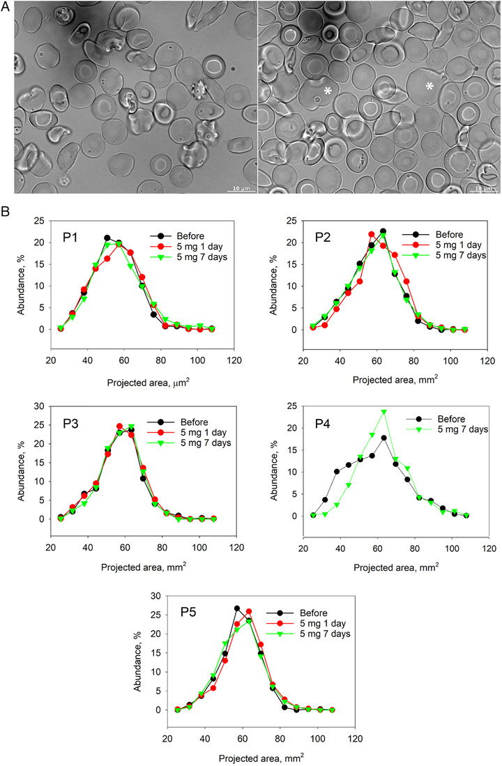
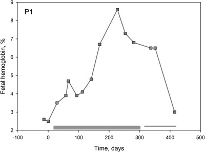
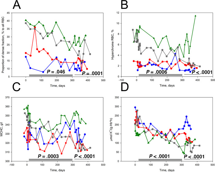
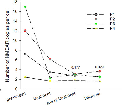
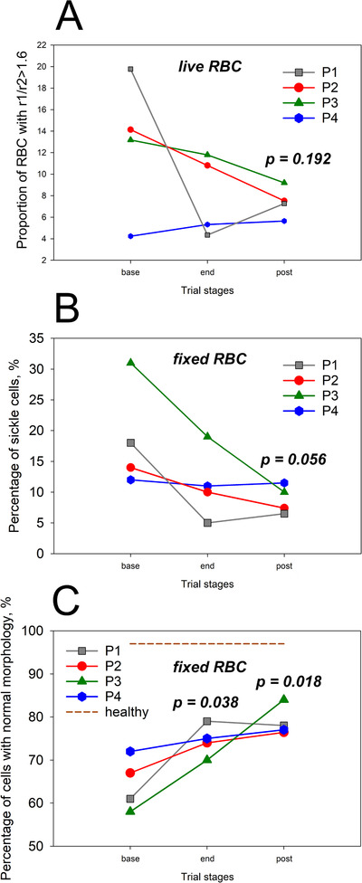
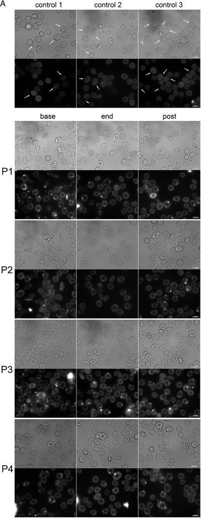
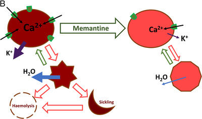
An increase in abundance and activity of N-methyl D-aspartate receptors (NMDAR) was previously reported for red blood cells (RBCs) of sickle cell disease (SCD) patients. Increased Ca2+ uptake through the receptor supported dehydration and RBC damage. In a pilot phase IIa-b clinical trial MemSID, memantine, a blocker of NMDAR, was used for treatment of four patients for 12 months. Two more patients that have enrolled into the study did not finish it. One of them had psychotic event following the involuntary overdose of the drug, whereas the other had vertigo and could not comply to the trial visits schedule. Acute and durable responses of RBCs of SCD patients to daily oral administration of memantine were monitored. Markers of RBC turnover, changes in cell density, and alterations in ion handling and RBC morphology were assessed. Acute transient shifts in intracellular Ca2+, volume and density, and reduction in plasma lactate dehydrogenate activity were observed already within the first month of treatment. Durable effects of memantine included (a) decrease in reticulocyte counts, (b) reduction in reticulocyte hemoglobinization, (c) advanced membrane maturation and its stabilization as follows from reduction in the number of NMDAR per cell and reduction in hemolysis, and (iv) rehydration and decrease in K+ leakage from patients' RBC. Memantine therapy resulted in reduction in number of cells with sickle morphology that was sustained at least over 2 months after therapy was stopped indicating an improvement in RBC longevity.


Conflict of interest statement

The authors declare no conflict of interest. The University of Zurich holds the patent to use memantine against sickle cell disease.

Figures

FIGURE 1
FIGURE 1
Acute effects of memantine therapy. Plasma memantine levels for SCD patients 1‐6 during the up‐dose phase. # stands for 1 week after the memantine therapy was discontinued (A). Alterations in the intracellular‐free Ca2+ measured as fluo‐4 fluorescence intensity (B), MCHC (C), MCV and (D) in RBCs of SCD patients 1‐6 during the up‐dose phase. ECell abundance in high density fraction (E). Plasma lactate dehydrogenase (LDH) activity in patients 1‐6 during the up‐dose phase (F). LDH activity values showed a significant decline by the end of up‐dose period (paired t‐test was applied comparing the last to the first value)
FIGURE 2
FIGURE 2
Changes in RBC morphology in response to memantine treatment during the up‐dosing phase. Comparison of RBC morphology of patient P1 before the onset of treatment (left panel) and at the up‐dose phase (right panel) (A). Abnormally large cells are highlighted with the star. Distribution of the projected areas of cells at the onset of the study, during the first day of treatment with 5 mg memantine and after a week of administration of 5 mg memantine a day (B).
FIGURE 3
FIGURE 3
Impact of durable memantine therapy on reticulocytes in patients 1‐4. Abundance of CD71+ reticulocytes (A), reticulocytes’ maturation state (CD71+/RNA+ cells; (B)) and their hemoglobinization (C) in SCD patients 1‐4 during the treatment, down‐dosing and follow‐up phases. D, Density of mature RBCs as a function of hemoglobin content of reticulocytes for cell of SCD patients on memantine trial. Bars indicate the treatment phase and the down‐dose and follow‐up periods. Significance of changes between the trial phases was assessed using a linear mixed model in which phases were chosen as variables and patients used as a random effect. The P‐values are shown when the treatment and down‐dose/follow‐up phases were compared with the pretreatment phase
FIGURE 4
FIGURE 4
Time‐course of changes in HbF abundance for patient P1 during the trial
FIGURE 5
FIGURE 5
Effect of memantine treatment on RBC hydration state and membrane maturation in patients 1‐4. Impact of durable memantine therapy on the abundance of dense cells (A) and hyperchrome cells (B), MCHC (C), and passive K+ leakage from RBCs (D). Significance of changes between the trial phases was assessed using a linear mixed model in which phases were chosen as variables and patients used as a random effect. The P‐values are shown when the treatment and down‐dose/follow‐up phases were compared with the pretreatment phase. Color‐coding for the P1‐P4 is similar to that in Fig. 3
FIGURE 6
FIGURE 6
Changes in abundance of NMDARs in mature RBCs as a result of memantine treatment. One‐way repeated measures ANOVA on ranks with was applied to estimate the difference in receptor abundance over the treatment phases (compared to the pretreatment phase). Numbers above the graphs are the p values
FIGURE 7
FIGURE 7
Morphological changes in RBCs upon memantine therapy in SCD patients 1‐4. Percentage of elongated cells (shortest to longest diameters R1/R2 > 1.6) observed for native cells using bright‐field microscopy (A). Percentage of fixed RBCs with sickled morphology (B). Abundance of fixed cells with normal morphology (C)
FIGURE 8
FIGURE 8
Calcium sequestration and the impact of Ca2+‐targeting memantine therapy on RBCs of SCD patients. (A) RBC morphology and the intracellular Ca2+ distribution in RBC of healthy donors (control1‐control3) and for patients P1‐P4 before the onset of treatment (base), by the end of treatment (end) and by the end of follow‐up phase (post). Arrows highlight nanovesicles filled with Ca2+ in cells of healthy patients. (B) Summary on the effect of memantine on RBC of SCD patients. Memantine therapy resulted in reduction in hemoglobin content in reticulocytes and RBCs, a decrease in the number of NMDARs per cell and improved hydration that was caused by a decline in K+ leak. As a result, irreversible sickling was avoided, and the longevity of RBCs improved
FIGURE 8
FIGURE 8
Calcium sequestration and the impact of Ca2+‐targeting memantine therapy on RBCs of SCD patients. (A) RBC morphology and the intracellular Ca2+ distribution in RBC of healthy donors (control1‐control3) and for patients P1‐P4 before the onset of treatment (base), by the end of treatment (end) and by the end of follow‐up phase (post). Arrows highlight nanovesicles filled with Ca2+ in cells of healthy patients. (B) Summary on the effect of memantine on RBC of SCD patients. Memantine therapy resulted in reduction in hemoglobin content in reticulocytes and RBCs, a decrease in the number of NMDARs per cell and improved hydration that was caused by a decline in K+ leak. As a result, irreversible sickling was avoided, and the longevity of RBCs improved

References

    1. GBD 2013 Mortality and Causes of Death Collaborators . Global, regional, and national age‐sex specific all‐cause and cause‐specific mortality for 240 causes of death, 1990–2013: a systematic analysis for the Global Burden of Disease Study 2013. Lancet. 2015;385:117–71. - PMC - PubMed
    1. Piel FB, Steinberg MH, Rees DC. Sickle cell disease. N Engl J Med. 2017;377:305. - PubMed
    1. Rees DC, Williams TN, Gladwin MT. Sickle‐cell disease. Lancet. 2010;376:2018–31. - PubMed
    1. Bender MA. Sickle cell disease. In: GeneReviews(R) (ed. by Adam MP, Ardinger HH,Pagon RA, Wallace SE, Bean LJH, Stephens K & Amemiya A), Seattle, WA: University of Washington; 1993. - PubMed
    1. Ware RE, de Montalembert M, Tshilolo L, Abboud MR. Sickle cell disease. Lancet. 2017;390:311–23. - PubMed