

Somatotopic Mapping of the Fingers in the Somatosensory Cortex Using Functional Magnetic Resonance Imaging: A Review of Literature
- PMID: 35847829
- PMCID: PMC9277538
- DOI: 10.3389/fnana.2022.866848
Somatotopic Mapping of the Fingers in the Somatosensory Cortex Using Functional Magnetic Resonance Imaging: A Review of Literature
Abstract
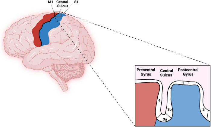
Multiple studies have demonstrated finger somatotopy in humans and other primates using a variety of brain mapping techniques including functional magnetic resonance imaging (fMRI). Here, we review the literature to better understand the reliability of fMRI for mapping the somatosensory cortex. We have chosen to focus on the hand and fingers as these areas have the largest representation and have been the subject of the largest number of somatotopic mapping experiments. Regardless of the methods used, individual finger somatosensory maps were found to be organized across Brodmann areas (BAs) 3b, 1, and 2 in lateral-to-medial and inferior-to-superior fashion moving from the thumb to the pinky. However, some consistent discrepancies are found that depend principally on the method used to stimulate the hand and fingers. Therefore, we suggest that a comparative analysis of different types of stimulation be performed to address the differences described in this review.
Keywords: cortical magnification; digit distance; digit overlap; fMRI; finger somatotopy; neuroimaging; somatosensory cortex.
Copyright © 2022 Janko, Thoenes, Park, Willoughby, Horton and Bolding.
Conflict of interest statement
The authors declare that the research was conducted in the absence of any commercial or financial relationships that could be construed as a potential conflict of interest.
Figures
References
-
- Al-Chalabi M., Reddy V., Alsalman I. (2022). Neuroanatomy, Posterior Column (dorsal column). StatPearls. Treasure Island, FL: StatPearls Publishing. - PubMed
Publication types
LinkOut - more resources
Full Text Sources