The Molecular Role of HIF1α Is Elucidated in Chronic Myeloid Leukemia
- PMID: 35847841
- PMCID: PMC9279726
- DOI: 10.3389/fonc.2022.912942
The Molecular Role of HIF1α Is Elucidated in Chronic Myeloid Leukemia
Abstract
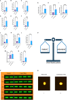
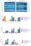
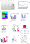
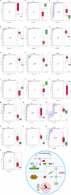
Chronic myeloid leukemia (CML) is potentially fatal blood cancer, but there is an unmet need to discover novel molecular biomarkers. The hypothesis of this study aimed to elucidate the relationship of HIF1α with the redox system, Krebs cycles, notch1, and other regulatory proteins to better understand the pathophysiology and clinical relevance in chronic myeloid leukemia (CML) patients, as the molecular mechanism of this axis is still not clear. This study included CML patient samples (n = 60; 60: blood; 10: bone marrow tissues) and compared them with healthy controls (n = 20; blood). Clinical diagnosis confirmed on bone marrow aspiration, marrow trephine biopsy, and BCR/ABL1 translocation. Cases were subclassified into chronic, accelerated, and blast crises as per WHO guidelines. Molecular experiments included redox parameters, DNA fragmentation, Krebs cycle metabolites, and gene expression by RT-PCR/Western blot/LC-MS, PPI (STRING), Pearson correlation, and ROC curve analysis. Here, our findings show that p210/p190BCR/ABL1 translocation is common in all blast crisis phases of CML. Redox factor/Krebs oncometabolite concentrations were high, leading to upregulation and stabilization of HIF1α. HIF1α leads to the pathogenesis in CML cells by upregulating their downstream genes (Notch 2/4/Ikaros/SIRT1/Foxo-3a/p53, etc.). Whereas, downregulated ubiquitin proteasomal and apoptotic factors in CML pateints, can trigger degradation of HIF1α through proline hydroxylation. However, HIF1α showed a negative corelation with the notch1 pathway. Notch1 plays a tumor-suppressive role in CML and might have the potential to be used as a diagnostic marker along with other factors in CML patients. The outcome also revealed that oxidant treatment could not be effective in augmentation with conventional therapy because CML cells can enhance the levels of antioxidants for their survival. HIF1α might be a novel therapeutic target other than BCR/ABL1 translocation.
Keywords: BCR/ABL1 translocation; HIF1α; Krebs metabolite; chronic myeloid leukemia (CML); hypoxia; notch signaling pathway; redox system.
Copyright © 2022 Singh, Singh, Kushwaha, Verma, Tripathi and Mahdi.
Conflict of interest statement
The authors declare that the research was conducted in the absence of any commercial or financial relationships that could be construed as a potential conflict of interest.
Figures


Similar articles
-
The bioengineered HALOA complex induces anoikis in chronic myeloid leukemia cells by targeting the BCR-ABL/Notch/Ikaros/Redox/Inflammation axis.J Med Life. 2022 May;15(5):606-616. doi: 10.25122/jml-2021-0230. J Med Life. 2022. PMID: 35815090 Free PMC article.
-
Chronic Myeloid Leukemia with cryptic Philadelphia translocation and extramedullary B-lymphoid blast phase as an initial presentation.Acta Biomed. 2018 Apr 3;89(3-S):38-44. doi: 10.23750/abm.v89i3-S.7219. Acta Biomed. 2018. PMID: 29633732 Free PMC article.
-
miR-96 acts as a tumor suppressor via targeting the BCR-ABL1 oncogene in chronic myeloid leukemia blastic transformation.Biomed Pharmacother. 2019 Nov;119:109413. doi: 10.1016/j.biopha.2019.109413. Epub 2019 Sep 10. Biomed Pharmacother. 2019. PMID: 31518872
-
New Developments in Chronic Myeloid Leukemia: Implications for Therapy.Iran J Cancer Prev. 2016 Feb 22;9(1):e3961. doi: 10.17795/ijcp-3961. eCollection 2016 Feb. Iran J Cancer Prev. 2016. PMID: 27366312 Free PMC article. Review.
-
Reactive oxygen species in BCR-ABL1-expressing cells - relevance to chronic myeloid leukemia.Acta Biochim Pol. 2017;64(1):1-10. doi: 10.18388/abp.2016_1396. Epub 2016 Dec 1. Acta Biochim Pol. 2017. PMID: 27904889 Review.
Cited by
-
Exploring novel protein biomarkers for early-stage diagnosis and prognosis of T-acute lymphoblastic leukemia (T-ALL).Hematol Transfus Cell Ther. 2024 Dec;46 Suppl 6(Suppl 6):S93-S111. doi: 10.1016/j.htct.2024.02.016. Epub 2024 Mar 28. Hematol Transfus Cell Ther. 2024. PMID: 38584071 Free PMC article.
-
Next-generation leukemia diagnostics: Integrating LC-MS/MS proteomics with liquid biopsy platforms.J Liq Biopsy. 2025 Aug 8;9:100324. doi: 10.1016/j.jlb.2025.100324. eCollection 2025 Sep. J Liq Biopsy. 2025. PMID: 40831732 Free PMC article. Review.
-
Regulation of Hypoxia Dependent Reprogramming of Cancer Metabolism: Role of HIF-1 and Its Potential Therapeutic Implications in Leukemia.Asian Pac J Cancer Prev. 2024 Apr 1;25(4):1121-1134. doi: 10.31557/APJCP.2024.25.4.1121. Asian Pac J Cancer Prev. 2024. PMID: 38679971 Free PMC article. Review.
-
The Bone Marrow Immune Microenvironment in CML: Treatment Responses, Treatment-Free Remission, and Therapeutic Vulnerabilities.Curr Hematol Malig Rep. 2023 Apr;18(2):19-32. doi: 10.1007/s11899-023-00688-6. Epub 2023 Feb 13. Curr Hematol Malig Rep. 2023. PMID: 36780103 Free PMC article. Review.
-
Breaking Barriers: The Role of the Bone Marrow Microenvironment in Multiple Myeloma Progression.Int J Mol Sci. 2025 Jul 28;26(15):7301. doi: 10.3390/ijms26157301. Int J Mol Sci. 2025. PMID: 40806429 Free PMC article. Review.
References
LinkOut - more resources
Full Text Sources
Research Materials
Miscellaneous

