. 2022 Jun 29:12:908603.
doi: 10.3389/fonc.2022.908603. eCollection 2022.

Proteomic Analysis Identifies p62/SQSTM1 as a Critical Player in PARP Inhibitor Resistance

Affiliations

Proteomic Analysis Identifies p62/SQSTM1 as a Critical Player in PARP Inhibitor Resistance

Mohammed Hafiz Uddin et al. Front Oncol. .

Abstract

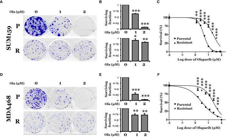
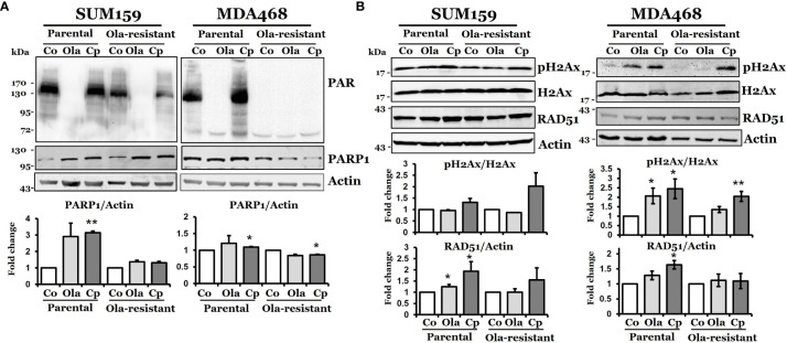
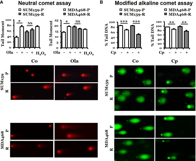
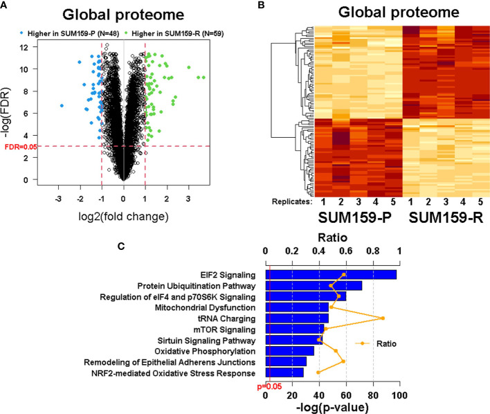
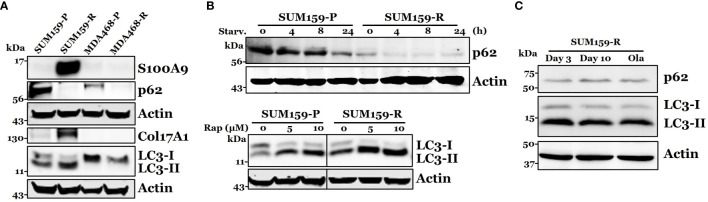
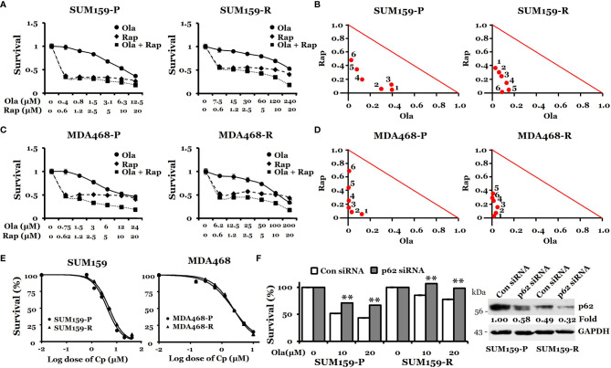
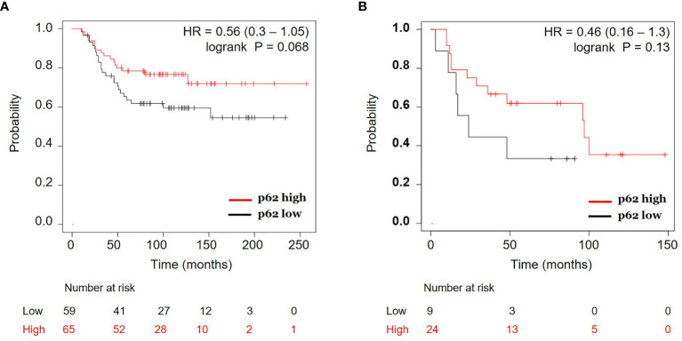
Poly (ADP-ribose) polymerase (PARP) inhibitors (PARPis) are currently being used for treating breast cancer patients with deleterious or suspected deleterious germline BRCA-mutated, HER2-negative locally advanced or metastatic diseases. Despite durable responses, almost all patients receiving PARPis ultimately develop resistance and succumb to their illness, but the mechanism of PARPi resistance is not fully understood. To better understand the mechanism of PARPi resistance, we established two olaparib-resistant SUM159 and MDA468 cells by chronically exposing olaparib-sensitive SUM159 and MDA468 cells to olaparib. Olaparib-resistant SUM159 and MDA468 cells displayed 5-fold and 7-fold more resistance over their corresponding counterparts. Despite defects in PARPi-induced DNA damage, these olaparib-resistant cells are sensitive to cisplatin-induced cell death. Using an unbiased proteomic approach, we identified 6 447 proteins, of which 107 proteins were differentially expressed between olaparib-sensitive and -resistant cells. Ingenuity pathway analysis (IPA) revealed a number of pathways that are significantly altered, including mTOR and ubiquitin pathways. Among these differentially expressed proteins, p62/SQSTM1 (thereafter p62), a scaffold protein, plays a critical role in binding to and delivering the ubiquitinated proteins to the autophagosome membrane for autophagic degradation, was significantly downregulated in olaparib-resistant cells. We found that autophagy inducers rapamycin and everolimus synergistically sensitize olaparib-resistant cells to olaparib. Moreover, p62 protein expression was correlated with better overall survival in estrogen receptor-negative breast cancer. Thus, these findings suggest that PARPi-sensitive TNBC cells hyperactivate autophagy as they develop acquired resistance and that pharmacological stimulation of excessive autophagy could lead to cell death and thus overcome PARPi resistance.

Keywords: PARP inhibitor; TNBC; autophagy; p62/SQSTM1; proteomics; rapamycin; resistance.


Conflict of interest statement

The authors declare that the research was conducted in the absence of any commercial or financial relationships that could be construed as a potential conflict of interest.

Figures

Figure 1
Figure 1
Clonogenic survival of PARP inhibitor olaparib-sensitive and -resistant TNBC cells. (A, D) Colony formation assay for SUM159 and MDA468 parental (P) and olaparib resistant (R) cells. Cells were treated with either DMSO or olaparib for 72 hrs and allowed to grow in a drug-free medium for 10 days. All treatments were carried out in triplicate, and the images are representative of the response to olaparib. (B, E) Surviving fraction calculated from A and D, respectively. (C, F) Dose-response curve for parental and olaparib resistant SUM159 and MDA468 cells. Resistant cells were obtained by selection with increasing concentrations of olaparib in the culture media for six months. After the development of resistance, SUM159-R and MDA468-R cells were maintained in media containing 25 µM and 15 µM olaparib, respectively. Cell viability was measured using MTT assay after treating with varying concentrations of olaparib for 72 hrs. After adding MTT to the media, cells were incubated at 37˚C for 2 hrs. The formazan crystals were dissolved in DMSO and then read by spectrophotometer at 570 nm. Data represented as mean ± standard deviation (SD). All experiments were done in triplicates. Ola, olaparib. Student’s t-test: *p <0.05; **P <0.01 and ***P <0.001.
Figure 2
Figure 2
DNA damage response in olaparib-sensitive and -resistant TNBC cells. Western blot analysis of DNA damage response associated proteins, PAR, PARP-1 (A), pH2AX, and RAD51 (B) in parental and olaparib resistant SUM159 and MDA468 cells. Cells were treated with 5 µM olaparib or 2 µM cisplatin for 24 hrs. SUM159-R and MDA468-R cells were maintained in drug-free media 2-3 days before the experiment. For the determination of band density, NIH ImageJ 1.5Oi software was used. Band densities were normalized against Actin or total H2AX. Images are representative of three independent experiments. Co, control; Ola, olaparib and Cp, cisplatin. Student’s t-test: *p < 0.05 and **p < 0.01.
Figure 3
Figure 3
Determination of DNA damage response in parental and olaparib resistant SUM159 and MDA468 cells by comet assays. (A) Neutral comet assay for detecting double-strand breaks in DNA in the form of tail moment. Parental and olaparib resistant SUM159 and MDA468 cells were treated with 15 µM and 10 µM olaparib for 24 hrs, respectively, followed by the electrophoresis of nuclear DNA. An average of 50 cells was used to calculate the tail moments. The lower panel shows a representative image of each group. (B) Modified alkaline comet assay for detecting DNA crosslinks in the form of percent tail DNA. Parental and olaparib resistant SUM159 and MDA468 cells were treated with 2 µM cisplatin for 24 hrs before the electrophoresis of nuclear DNA. An average of 50 cells was used to calculate the percent tail DNA. The lower panel shows a representative image from each group. Co, control; Ola, olaparib; Cp, cisplatin; P, parental; R, resistant and NS, not significant. Student’s t-test: *p < 0.05; **p < 0.01 and ***p < 0.001.
Figure 4
Figure 4
Quantitative measurement of the global proteome using TMT labeling in SUM159 parental and resistant TNBC cells. (A) Volcano plots showing differentially expressed proteins in SUM159-R cells compared to SUM159-P cells. Each dot represents one protein. Blue and green dots represent proteins that are significantly higher in SUM159-P cells (N = 48) and those that are significantly higher in SUM159-R cells (N = 59), respectively, with FDR<0.05 and fold change (FC) ≥ 2. (B) Heatmap generated from proteins detected in all samples by hierarchical clustering. FDR<0.05 and FC ≥2. (C) Top 10 canonical pathways related to olaparib resistance derived from the global proteome’s ingenuity pathway analysis (IPA). P, parental and R, resistant.
Figure 5
Figure 5
Confirmation of S100A9, p62, COL17A1, and LC3 expression in olaparib resistant cells. (A) Western blot analysis of S100A9, p62, COL17A1, and p62 associated protein LC3-I, II in both parental and resistant SUM159 and MDA468 cells. (B) Effect of autophagy stimulation on p62 and LC3 expression. Parental and resistant SUM159 cells were starved for indicated periods (upper panel) or treated with two different doses (5 µM and 10 µM) of rapamycin for 24 hrs (lower panel). The levels of p62 or LC3 were measured by western blotting. Rap, rapamycin and starv, starvation. (C) Effect of olaparib withdrawal on LC3 and p62 levels. SUM159-R cells were maintained in olaparib-free media for 3 days and 10 days or in olaparib-containing (25 µM) medium (ola).). P, parental; R, resistant; Starv., starvation; Rap, rapamycin; and Ola, olaparib.
Figure 6
Figure 6
Effects of rapamycin plus olaparib or cisplatin alone on the growth of olaparib-resistant TNBC cells. (A, C), Synergistic effects of rapamycin and olaparib treatment on the growth of SUM159-P and SUM159-R cells (A) and MDA468-P and MDA468-R cells (C). Cells were treated with indicated drug combination for 72 hrs. (B, D), Normalized isobologram for SUM159-P and SUM159-R cells (B) and MDA468-P and MDA468-R cells (D). (E) Sensitivity of olaparib-resistant cells to cisplatin in parental and olaparib resistant SUM159 cells (left) and MDA468 cells (right). Cell viability was measured using MTT assay after treatment with varying concentrations of cisplatin for 72 hrs. All experiments were performed in triplicates. P, parental; R, resistant; Ola, olaparib; Cp, cisplatin; Rap, rapamycin and con, control. (F) Effect of p62 knockdown on olaparib sensitivity in SUM159 cells. SUM159-P and SUM159-R cells were transfected with p62 siRNA or non-target siRNA (Con siRNA). MTT was performed to assess olaparib sensitivity (left panel), and the level of p62 knockdown was evaluated by western blotting (right panel). Student’s t-test: **p < 0.01.
Figure 7
Figure 7
Correlation of p62 protein expression and overall survival in estrogen receptor (ER) negative breast cancer. Kaplan-Meier curves of ER-negative breast cancer patients with p62 protein high vs. p62 protein low for overall survival were generated using the webtool Kaplan-Meier Plotter (http://kmplot.com) (24) using two breast cancer datasets: (A) Liu et al. (2014) (27) and (B) Tang et al. (2018) (28). HR, hazard ratio.

Similar articles

Cited by

References

    1. Perou CM, Sorlie T, Eisen MB, van de Rijn M, Jeffrey SS, Rees CA, et al. . Molecular Portraits of Human Breast Tumours. Nature (2000) 406(6797):747–52. doi: 10.1038/35021093 - DOI - PubMed
    1. Dent R, Trudeau M, Pritchard KI, Hanna WM, Kahn HK, Sawka CA, et al. . Triple-Negative Breast Cancer: Clinical Features and Patterns of Recurrence. Clin Cancer Res (2007) 13(15 Pt 1):4429–34. doi: 10.1158/1078-0432.CCR-06-3045 - DOI - PubMed
    1. Perez EA, Moreno-Aspitia A, Aubrey Thompson E, Andorfer CA. Adjuvant Therapy of Triple Negative Breast Cancer. Breast Cancer Res Treat (2010) 120(2):285–91. doi: 10.1007/s10549-010-0736-z - DOI - PMC - PubMed
    1. Kang SP, Martel M, Harris LN. Triple Negative Breast Cancer: Current Understanding of Biology and Treatment Options. Curr Opin Obstet Gynecol (2008) 20(1):40–6. doi: 10.1097/GCO.0b013e3282f40de9 - DOI - PubMed
    1. Isakoff SJ. Triple-Negative Breast Cancer: Role of Specific Chemotherapy Agents. Cancer J (2010) 16(1):53–61. doi: 10.1097/PPO.0b013e3181d24ff7 - DOI - PMC - PubMed