Review
. 2022 Jun 29:9:923794.
doi: 10.3389/fmolb.2022.923794. eCollection 2022.

Librational Dynamics of Spin-Labeled Membranes at Cryogenic Temperatures From Echo-Detected ED-EPR Spectra

Affiliations
Review

Librational Dynamics of Spin-Labeled Membranes at Cryogenic Temperatures From Echo-Detected ED-EPR Spectra

Rosa Bartucci et al. Front Mol Biosci. .

Abstract

Methods of electron spin echo of pulse electron paramagnetic resonance (EPR) spectroscopy are increasingly employed to investigate biophysical properties of nitroxide-labeled biosystems at cryogenic temperatures. Two-pulse echo-detected ED-spectra have proven to be valuable tools to describe the librational dynamics in the low-temperature phases of both lipids and proteins in membranes. The motional parameter, α 2 τ C , given by the product of the mean-square angular amplitude, α 2 , and the rotational correlation time, τ C , of the motion, is readily determined from the nitroxide ED-spectra as well as from the W-relaxation rate curves. An independent evaluation of α 2 is obtained from the motionally averaged 14N-hyperfine splitting separation in the continuous wave cw-EPR spectra. Finally, the rotational correlation time τ C can be estimated by combining ED- and cw-EPR data. In this mini-review, results on the librational dynamics in model and natural membranes are illustrated.

Keywords: Na, K-ATPase; echo-detected ED-spectra; electron paramagnetic resonance; electron spin echo; librations; model membranes; spin label.


Conflict of interest statement

The authors declare that the research was conducted in the absence of any commercial or financial relationships that could be construed as a potential conflict of interest.

Figures
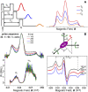
FIGURE 1
FIGURE 1
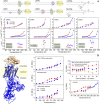
(A) Two-pulse primary echo sequences and echo amplitudes decrease with increasing the interpulse delay time, τ; simulated examples of corresponding echo-detected ED-spectra of chain-labeled nitroxide in membranes. (B) Two-pulse (π/2-τ-π with microwave pulse widths of 32 and 64 ns) ED-EPR spectra of 5-PCSL in DPPC bilayers at T = 200 K recorded at incremented interpulse spacings τ (from top to bottom). Solid lines are the normalized experimental spectra, and dashed lines are simulations for isotropic librational motion. Underneath are reported the anisotropic part of the relaxation rate, W-spectra, obtained according to Eq. 1 from pairs of spectra with interpulse separations of τ 1 = 168 and τ 2 = 296 ns, τ 1 = 168 ns and τ 2 = 424 ns, or τ 1 = 168 ns and τ 2 = 552 ns. Schematic illustration of isotropic librational motion: the nitroxide molecule performs oscillations of small angular amplitude, α, about the three nitroxide axes. cw-EPR spectra of 5-PCSL in DPPC bilayers at 150, 220, and 260 K. ED-, W-, and cw-EPR spectra are taken from Aloi et al. (2017).
FIGURE 2
FIGURE 2
(A) Chemical structure of the lipids DPPC, DHPC, POPC, and DOPC and of the chain-labeled phosphatidylcholine spin-label 5-PCSL and 16-PCSL. Characterization of the segmental librational motion in DPPC, DHPC, POPC, and DOPC membranes spin-labeled with 5- and 16-PCSL via the temperature dependence of the (i) amplitude-correlation time product, α2τC , (ii) mean-square angular amplitude, α2 , and (iii) correlation time, τC . Error bars for α2 are within the symbols. Data for DPPC and DHPC are adapted from Aloi et al. (2017), and those for POPC and DOPC are from Aloi et al. (2019). (B) Na,K-ATPase membrane: crystal structure of the enzyme (PDB ID 4RES (Laursen et al., 2015)) and schematic bilayer region. Temperature dependence of the relaxation rate W L for 5- and 14-SASL in the Na,K-ATPase membrane, in bilayers of extracted lipids and at the lipid–protein interface. Chain positional profile, that is, W L vs. n, at T = 180 K of n-SASL in Na,K-ATPase membranes, in bilayers of the extracted lipids and at the lipid–protein interface. Temperature dependence of W L for 14-SASL in bilayers of extracted lipids and at the lipid–protein interface and for 5-MSL in the Na,K-ATPase protein. Data are adapted from Guzzi et al. (2015).

Similar articles

References

    1. Aloi E., Bartucci R. (2022). Influence of Hydration on Segmental Chain Librations and Dynamical Transition in Lipid Bilayers. Biochimica Biophysica Acta (BBA) - Biomembr. 1864, 183805. 10.1016/j.bbamem.2021.183805 - DOI - PubMed
    1. Aloi E., Bartucci R. (2019). Interdigitated Lamellar Phases in the Frozen State: Spin-Label CW- and FT-EPR. Biophys. Chem. 253, 106229. 10.1016/j.bpc.2019.106229 - DOI - PubMed
    1. Aloi E., Guo J. -H., Guzzi R., Jiang R. -W., Ladefoged L. K., Marsh D., et al. (2021). Geometry and Water Accessibility of the Inhibitor Binding Site of Na+-Pump: Pulse- and CW-EPR Study. Biophys. J. 120, 2679–2690. - PMC - PubMed
    1. Aloi E., Guzzi R., Bartucci R. (2019). Unsaturated Lipid Bilayers at Cryogenic Temperature: Librational Dynamics of Chain-Labeled Lipids from Pulsed and CW-EPR. Phys. Chem. Chem. Phys. 21, 18699–18705. 10.1039/c9cp03318a - DOI - PubMed
    1. Aloi E., Oranges M., Guzzi R., Bartucci R. (2017). Low-Temperature Dynamics of Chain-Labeled Lipids in Ester- and Ether-Linked Phosphatidylcholine Membranes. J. Phys. Chem. B 121, 9239–9246. 10.1021/acs.jpcb.7b07386 - DOI - PubMed

LinkOut - more resources