

Monodispersed Sirolimus-Loaded PLGA Microspheres with a Controlled Degree of Drug-Polymer Phase Separation for Drug-Coated Implantable Medical Devices and Subcutaneous Injection
- PMID: 35848106
- PMCID: PMC9382632
- DOI: 10.1021/acsabm.2c00319
Monodispersed Sirolimus-Loaded PLGA Microspheres with a Controlled Degree of Drug-Polymer Phase Separation for Drug-Coated Implantable Medical Devices and Subcutaneous Injection
Abstract
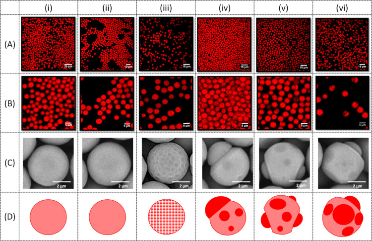
Monodispersed sirolimus (SRL)-loaded poly(lactic-co-glycolic acid) microspheres with a diameter of 1.8, 3.8, and 8.5 μm were produced by high-throughput microfluidic step emulsification─solvent evaporation using single crystal silicon chips consisted of 540-1710 terraced microchannels with a depth of 2, 4, or 5 μm arranged in 10 parallel arrays. Uniform sized droplets were generated over 25 h across all channels. Nearly 15% of the total drug was released by the initial burst release during an accelerated drug release testing performed at 37 °C using a hydrotropic solution containing 5.8 M N,N-diethylnicotinamide. After 24 h, 71% of the drug was still entrapped in the particles. The internal morphology of microspheres was investigated by fluorescence microscopy using Nile red as a selective fluorescent stain with higher binding affinity toward SRL. By increasing the drug loading from 33 to 50 wt %, the particle morphology evolved from homogeneous microspheres, in which the drug and polymer were perfectly mixed, to patchy particles, with amorphous drug patches embedded within a polymer matrix to anisotropic patchy Janus particles. Janus particles with fully segregated drug and polymer regions were achieved by pre-saturating the aqueous phase with the organic solvent, which decreased the rate of solvent evaporation and allowed enough time for complete phase separation. This approach to manufacturing drug-loaded monodisperse microparticles can enable the development of more effective implantable drug-delivery devices and improved methods for subcutaneous drug administration, which can lead to better therapeutic treatments.
Keywords: biodegradable polymer; controlled drug release; drug delivery; drug-eluting medical devices; poly(lactic-co-glycolic acid); step microfluidic emulsification.
Conflict of interest statement
The authors declare no competing financial interest.
Figures


Similar articles
-
Structured Biodegradable Polymeric Microparticles for Drug Delivery Produced Using Flow Focusing Glass Microfluidic Devices.ACS Appl Mater Interfaces. 2015 Oct 21;7(41):23132-43. doi: 10.1021/acsami.5b06943. Epub 2015 Oct 12. ACS Appl Mater Interfaces. 2015. PMID: 26423218
-
Effect of solvent on drug release and a spray-coated matrix of a sirolimus-eluting stent coated with poly(lactic-co-glycolic acid).Langmuir. 2014 Aug 26;30(33):10098-106. doi: 10.1021/la500452h. Epub 2014 Aug 13. Langmuir. 2014. PMID: 25090045
-
Influence of the microencapsulation method and peptide loading on poly(lactic acid) and poly(lactic-co-glycolic acid) degradation during in vitro testing.J Control Release. 1998 Feb 12;51(2-3):327-41. doi: 10.1016/s0168-3659(97)00188-0. J Control Release. 1998. PMID: 9685930
-
Ketoprofen-poly(D,L-lactic-co-glycolic acid) microspheres: influence of manufacturing parameters and type of polymer on the release characteristics.J Microencapsul. 1999 Jan-Feb;16(1):1-12. doi: 10.1080/026520499289266. J Microencapsul. 1999. PMID: 9972498
-
Poly(lactic-co-glycolic acid) devices: Production and applications for sustained protein delivery.Wiley Interdiscip Rev Nanomed Nanobiotechnol. 2018 Sep;10(5):e1516. doi: 10.1002/wnan.1516. Epub 2018 Mar 13. Wiley Interdiscip Rev Nanomed Nanobiotechnol. 2018. PMID: 29536634 Free PMC article. Review.
Cited by
-
PLGA-based long-acting injectable (LAI) formulations.J Control Release. 2025 Jun 10;382:113758. doi: 10.1016/j.jconrel.2025.113758. Epub 2025 Apr 21. J Control Release. 2025. PMID: 40268201 Review.
-
Everolimus-encapsulation in Pluronic P123 self-assembled micelles as drug delivery systems for drug-coated balloons.Int J Pharm X. 2024 Jan 10;7:100230. doi: 10.1016/j.ijpx.2024.100230. eCollection 2024 Jun. Int J Pharm X. 2024. PMID: 39668884 Free PMC article.
-
PLGA Implants for Controlled Drug Delivery and Regenerative Medicine: Advances, Challenges, and Clinical Potential.Pharmaceuticals (Basel). 2025 Apr 27;18(5):631. doi: 10.3390/ph18050631. Pharmaceuticals (Basel). 2025. PMID: 40430452 Free PMC article. Review.
-
Microstructure Formation and Characterization of Long-Acting Injectable Microspheres: The Gateway to Fully Controlled Drug Release Pattern.Int J Nanomedicine. 2024 Feb 19;19:1571-1595. doi: 10.2147/IJN.S445269. eCollection 2024. Int J Nanomedicine. 2024. PMID: 38406600 Free PMC article. Review.
-
Production of Hydrophobic Microparticles at Safe-To-Inject Sizes for Intravascular Administration.Pharmaceutics. 2025 Jan 6;17(1):64. doi: 10.3390/pharmaceutics17010064. Pharmaceutics. 2025. PMID: 39861712 Free PMC article.
References
Publication types
MeSH terms
Substances
LinkOut - more resources
Full Text Sources
Miscellaneous
