

Direct regulation of TCR rearrangement and expression by E proteins during early T cell development
- PMID: 35848146
- PMCID: PMC9669112
- DOI: 10.1002/wsbm.1578
Direct regulation of TCR rearrangement and expression by E proteins during early T cell development
Abstract
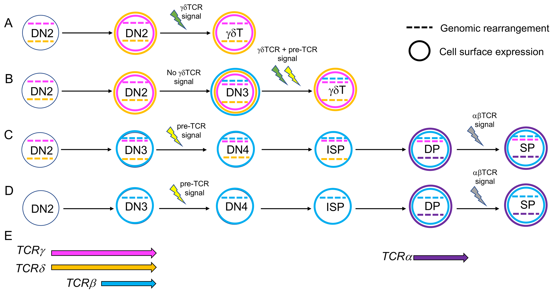
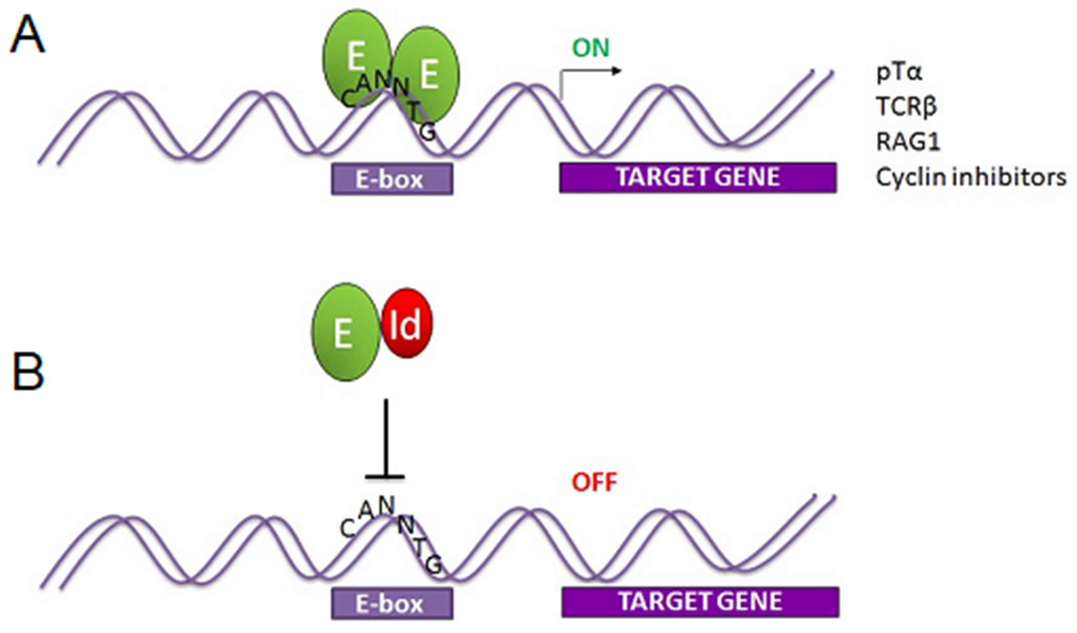
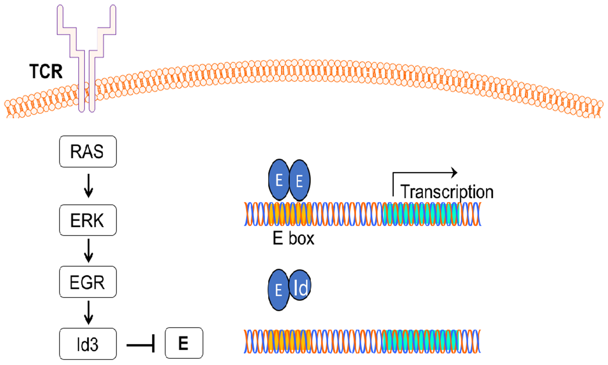
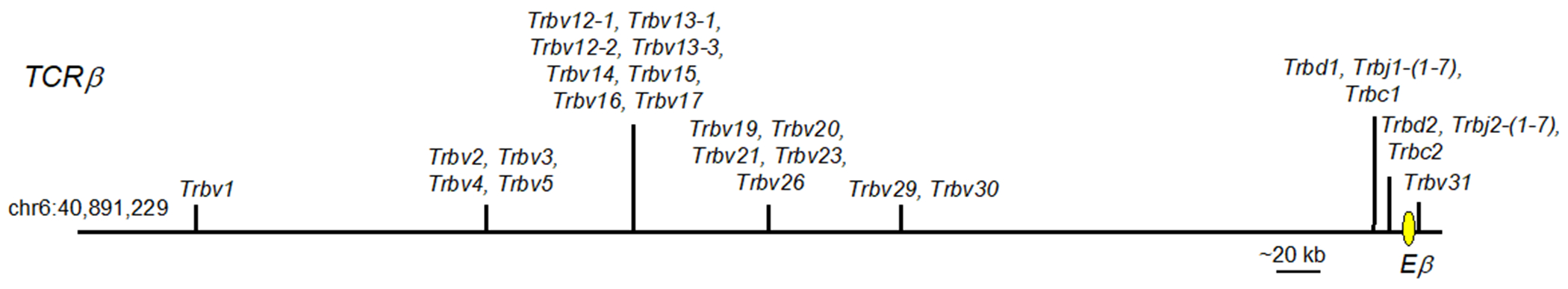
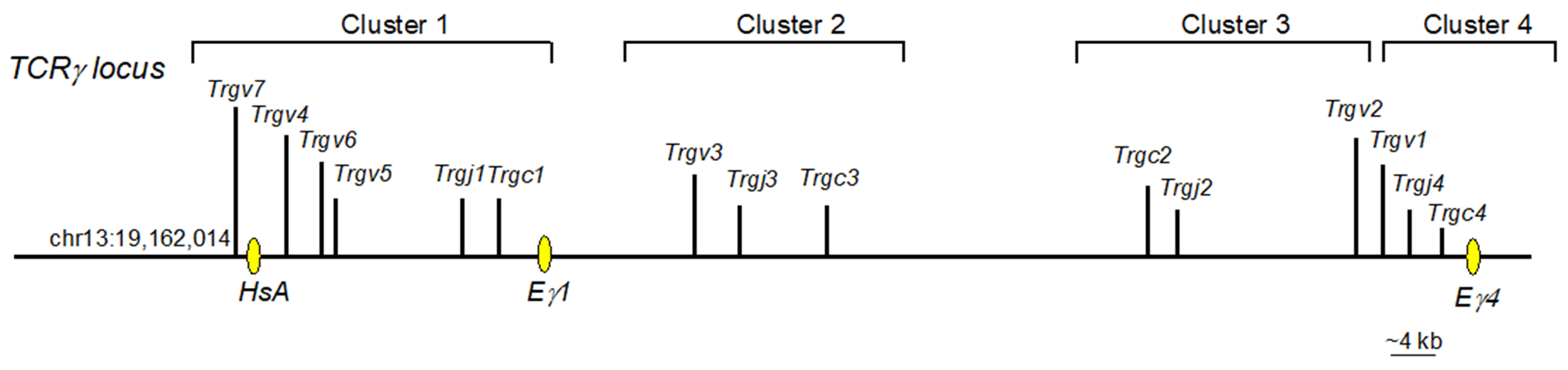
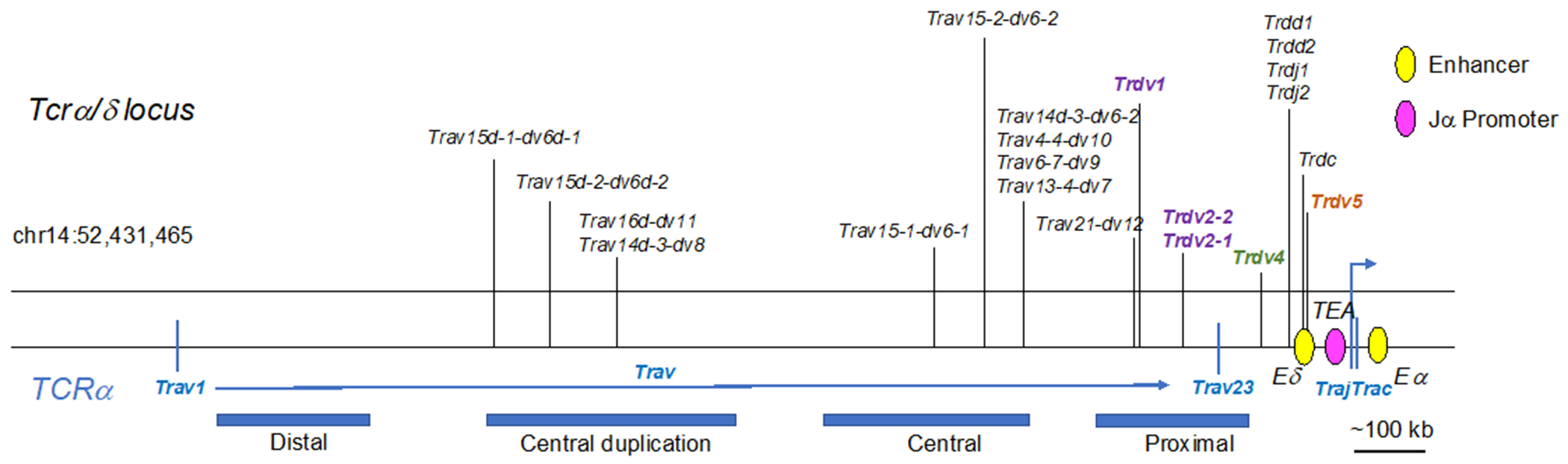
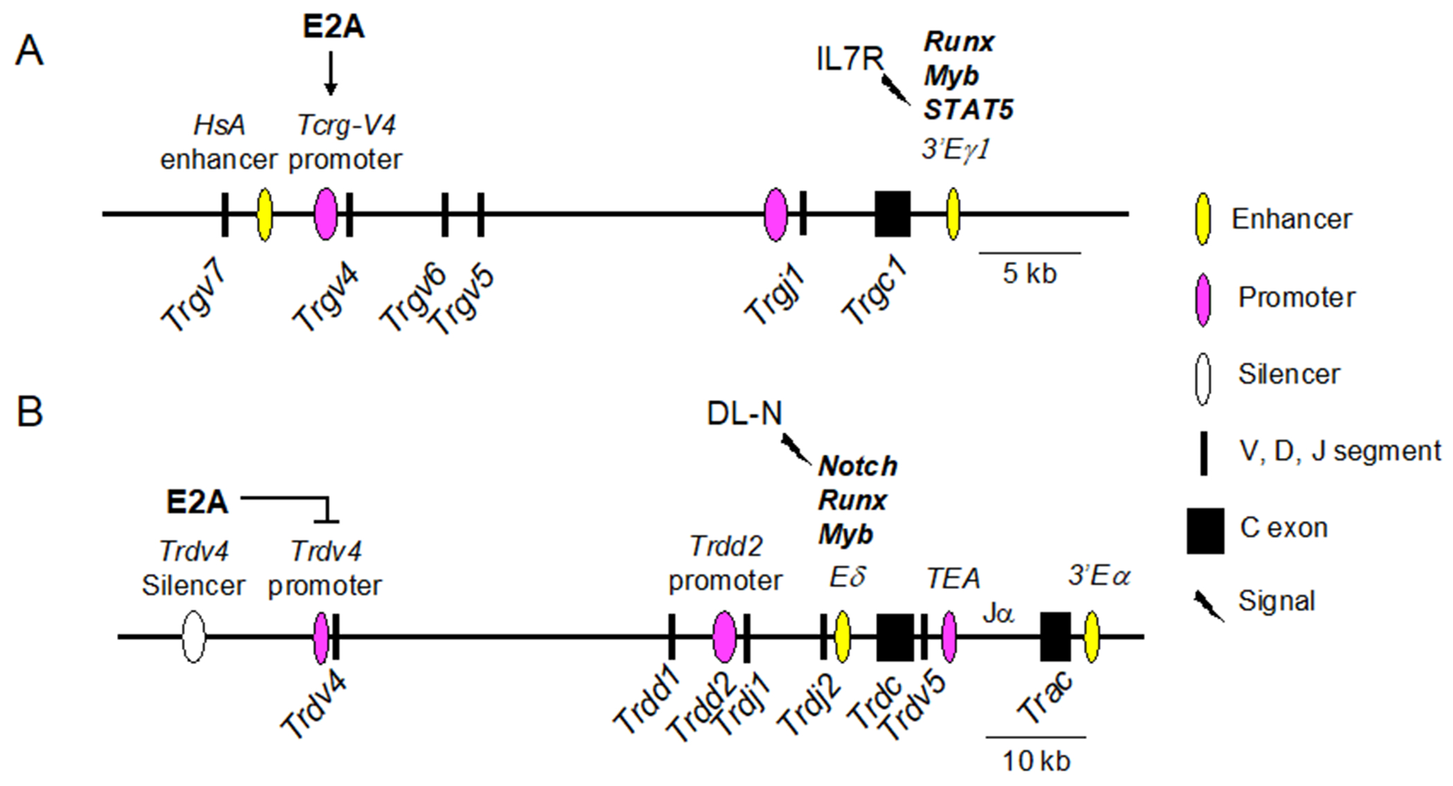
γδ T cells are widely distributed throughout mucosal and epithelial cell-rich tissues and are an important early source of IL-17 in response to several pathogens. Like αβ T cells, γδ T cells undergo a stepwise process of development in the thymus that requires recombination of genome-encoded segments to assemble mature T cell receptor (TCR) genes. This process is tightly controlled on multiple levels to enable TCR segment assembly while preventing the genomic instability inherent in the double-stranded DNA breaks that occur during this process. Each TCR locus has unique aspects in its structure and requirements, with different types of regulation before and after the αβ/γδ T cell fate choice. It has been known that Runx and Myb are critical transcriptional regulators of TCRγ and TCRδ expression, but the roles of E proteins in TCRγ and TCRδ regulation have been less well explored. Multiple lines of evidence show that E proteins are involved in TCR expression at many different levels, including the regulation of Rag recombinase gene expression and protein stability, induction of germline V segment expression, chromatin remodeling, and restriction of the fetal and adult γδTCR repertoires. Importantly, E proteins interact directly with the cis-regulatory elements of the TCRγ and TCRδ loci, controlling the predisposition of a cell to become an αβ T cell or a γδ T cell, even before the lineage-dictating TCR signaling events. This article is categorized under: Immune System Diseases > Stem Cells and Development Immune System Diseases > Genetics/Genomics/Epigenetics.
Keywords: E2A; T cell receptor; gamma delta T cell; gene rearrangement; thymus.
© 2022 Wiley Periodicals LLC.
Conflict of interest statement
Figures
Similar articles
-
Human alpha beta and gamma delta thymocyte development: TCR gene rearrangements, intracellular TCR beta expression, and gamma delta developmental potential--differences between men and mice.J Immunol. 2006 Feb 1;176(3):1543-52. doi: 10.4049/jimmunol.176.3.1543. J Immunol. 2006. PMID: 16424183 Free PMC article.
-
Deep sequencing of the human TCRγ and TCRβ repertoires suggests that TCRβ rearranges after αβ and γδ T cell commitment.Sci Transl Med. 2011 Jul 6;3(90):90ra61. doi: 10.1126/scitranslmed.3002536. Sci Transl Med. 2011. PMID: 21734177 Free PMC article.
-
Function of the TCR alpha enhancer in alphabeta and gammadelta T cells.Immunity. 1997 Oct;7(4):505-15. doi: 10.1016/s1074-7613(00)80372-6. Immunity. 1997. PMID: 9354471
-
αβ and γδ T cell receptors: Similar but different.J Leukoc Biol. 2020 Jun;107(6):1045-1055. doi: 10.1002/JLB.2MR1219-233R. Epub 2020 Jan 29. J Leukoc Biol. 2020. PMID: 31994778 Review.
-
Developmental regulation of V(D)J recombination at the TCR alpha/delta locus.Immunol Rev. 1998 Oct;165:131-47. doi: 10.1111/j.1600-065x.1998.tb01236.x. Immunol Rev. 1998. PMID: 9850858 Review.
Cited by
-
Interlinked roles for HEB and Id3 in fetal gamma-delta T cell commitment and functional programming.bioRxiv [Preprint]. 2025 Jun 8:2025.06.08.658490. doi: 10.1101/2025.06.08.658490. bioRxiv. 2025. PMID: 40501861 Free PMC article. Preprint.
-
E proteins control the development of NKγδT cells through their invariant T cell receptor.Nat Commun. 2024 Jun 13;15(1):5078. doi: 10.1038/s41467-024-49496-3. Nat Commun. 2024. PMID: 38871720 Free PMC article.
References
-
- Alonzo ES, Gottschalk RA, Das J, Egawa T, Hobbs RM, Pandolfi PP, … Sant’Angelo DB (2010). Development of promyelocytic zinc finger and ThPOK-expressing innate gamma delta T cells is controlled by strength of TCR signaling and Id3. J Immunol, 184(3), 1268–1279. doi:10.4049/jimmunol.0903218 - DOI - PMC - PubMed
Publication types
MeSH terms
Substances
Grants and funding
LinkOut - more resources
Full Text Sources
