. 2022 Jun 29:(184):10.3791/64040.
doi: 10.3791/64040.

Xenograft Skin Model to Manipulate Human Immune Responses In Vivo

Affiliations

Xenograft Skin Model to Manipulate Human Immune Responses In Vivo

Madison I Moss et al. J Vis Exp. .

Abstract

The human skin xenograft model, in which human donor skin is transplanted onto an immunodeficient mouse host, is an important option for translational research in skin immunology. Murine and human skin differ substantially in anatomy and immune cell composition. Therefore, traditional mouse models have limitations for dermatological research and drug discovery. However, successful xenotransplants are technically challenging and require optimal specimen and mouse graft site preparation for graft and host survival. The present protocol provides an optimized technique for transplanting human skin onto mice and discusses necessary considerations for downstream experimental aims. This report describes the appropriate preparation of a human donor skin sample, assembly of a surgical setup, mouse and surgical site preparation, skin transplantation, and post-surgical monitoring. Adherence to these methods allows for maintenance of xenografts for over 6 weeks post-surgery. The techniques outlined below allow maximum grafting efficiency due to the development of engineering controls, sterile technique, and pre- and post-surgical conditioning. Appropriate performance of the xenograft model results in long-lived human skin graft samples for experimental characterization of human skin and preclinical testing of compounds in vivo.


Figures

Figure 1:
Figure 1:. Method for bandaging mouse after surgery
The mouse is wrapped tightly in petrolatum gauze and transparent film dressing while under anesthesia. (A) Petrolatum gauze is placed over an area slightly larger than the surgical wound. (B) The transparent film dressing is prepared: a long strip is cut slightly wider than the petrolatum gauze. The backing covering the adhesive side of the transparent film dressing is partially removed. (C) The adhesive side of the film is firmly pressed against the back of the mouse, leaving an inch of space on the front end of the film. (D) The mouse is turned on its back. The overhanging front portion of the film is pressed onto the mouse, and the backing is removed. (E) The mouse is tightly wrapped in the film, and the remaining support is removed as the mouse is wrapped. (F) The mouse is successfully wrapped in the dressing, and its arms and legs are unconstrained.
Figure 2:
Figure 2:. Representative images from transplanted animals from Day 10 to 21 post-transplant
(A-C) Three different mice with optimal transplants at day 10. (D-E) Two different mice with transplants on day 21.
Figure 3:
Figure 3:. Immune cell chimerism in grafted skin
Flow cytometry data of human immune cell recovery from xenografts. Data is gated on the events of singlet, live, human CD45+ mouse CD45−. (A) Number of cells (Live Human CD45+ Events) recovered from two independent experiments. (B) Representative plots of human and mouse CD45+ immune cell staining in a successful graft (left) compared to graft with unsuccessful maintenance of the human immune compartment (right).
Figure 4:
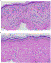
Figure 4:. Histology of grafted human skin at day 35 and 50
Grafted skin was harvested from mice on day 35 (A) or 50 (B), preserved in 10% formalin, and paraffin-embedded and stained for Hematoxylin and Eosin (H&E). (A) The epidermis shows moderate hyperplasia (acanthosis), slight hyper-granulosis, and compact orthokeratosis. In the papillary dermis, there are occasional dilated and congested capillaries. Melanocytes and melanin pigment are present in the basal layer of the epidermis. The dermis is moderately cellular and is composed of plump oval fibroblasts with pale syncytial cytoplasm and scattered lymphocytes. (B) The epidermis comprises stratified squamous epithelium, basket-weave orthokeratosis in the cornified layer, and is of normal thickness. Melanocytes and melanin pigment are present in the basal layer of the epidermis. The dermis shows oval fibroblasts and slightly enhanced extracellular matrix deposition.

References

    1. Zomer HD, Trentin AG Skin wound healing in humans and mice: Challenges in translational research. Journal of Dermatological Science. 90, (1), 3–12 (2018). - PubMed
    1. Wong VW, Sorkin M, Glotzbach JP, Longaker MT, Gurtner GC Surgical approaches to create murine models of human wound healing. Journal of Biomedicine & Biotechnology. 2011, 969618 (2011). - PMC - PubMed
    1. Mestas J, Hughes CCW Of mice and not men: differences between mouse and human immunology. The Journal of Immunology. 172, (5), 2731–2738 (2004). - PubMed
    1. Pasparakis M, Haase I, Nestle FO Mechanisms regulating skin immunity and inflammation. Nature Reviews Immunology. 14, (5), 289–301 (2014). - PubMed
    1. Sun H, Zhang Y-X, Li Y-M Generation of skin organoids: potential opportunities and challenges. Frontiers in Cell and Developmental Biology. 9, 3176 (2021). - PMC - PubMed

Publication types

LinkOut - more resources