

Luminal and Glandular Epithelial Cells from the Porcine Endometrium maintain Cell Type-Specific Marker Gene Expression in Air-Liquid Interface Culture
- PMID: 35849251
- PMCID: PMC9622560
- DOI: 10.1007/s12015-022-10410-3
Luminal and Glandular Epithelial Cells from the Porcine Endometrium maintain Cell Type-Specific Marker Gene Expression in Air-Liquid Interface Culture
Abstract
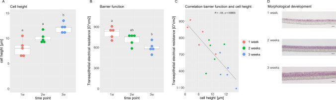
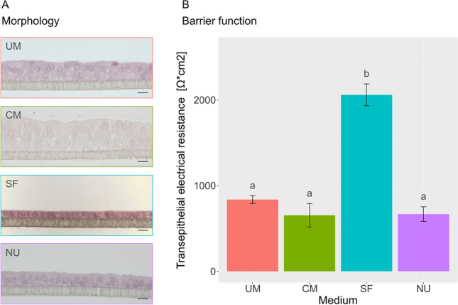
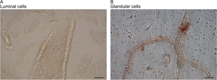
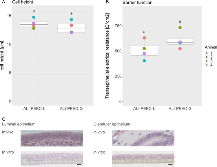
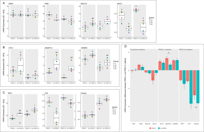
Two different types of epithelial cells constitute the inner surface of the endometrium. While luminal epithelial cells line the uterine cavity and build the embryo-maternal contact zone, glandular epithelial cells form tubular glands reaching deeply into the endometrial stroma. To facilitate investigations considering the functional and molecular differences between the two populations of epithelial cells and their contribution to reproductive processes, we aimed at establishing differentiated in vitro models of both the luminal and the glandular epithelium of the porcine endometrium using an air-liquid interface (ALI) approach. We first tested if porcine luminal endometrium epithelial cells (PEEC-L) reproducibly form differentiated epithelial monolayers under ALI conditions by monitoring the morphology and the trans-epithelial electrical resistance (TEER). Subsequently, luminal (PEEC-L) and glandular epithelial cells (PEEC-G) were consecutively isolated from the endometrium of the uterine horn. Both cell types were characterized by marker gene expression analysis immediately after isolation. Cells were separately grown at the ALI and assessed by means of histomorphometry, TEER, and marker gene expression after 3 weeks of culture. PEEC-L and PEEC-G formed polarized monolayers of differentiated epithelial cells with a moderate TEER and in vivo-like morphology at the ALI. They exhibited distinct patterns of functional and cell type-specific marker gene expression after isolation and largely maintained these patterns during the culture period. The here presented cell culture procedure for PEEC-L and -G offers new opportunities to study the impact of embryonic signals, endocrine effectors, and reproductive toxins on both porcine endometrial epithelial cell types under standardized in vitro conditions. Created with BioRender.com .
Keywords: Air–liquid interface; Cell culture; Endometrium; Glandular epithelium; Luminal epithelium.
© 2022. The Author(s).
Conflict of interest statement
The authors have no conflict of interest to declare.
Figures
Similar articles
-
Generation of Human Endometrial Assembloids with a Luminal Epithelium using Air-Liquid Interface Culture Methods.Adv Sci (Weinh). 2023 Oct;10(30):e2301868. doi: 10.1002/advs.202301868. Epub 2023 Aug 27. Adv Sci (Weinh). 2023. PMID: 37635169 Free PMC article.
-
Morphometric analysis of the uterine endometrium of swine on days 12 and 16 postestrus.Anat Rec A Discov Mol Cell Evol Biol. 2003 Jan;270(1):59-66. doi: 10.1002/ar.a.10182. Anat Rec A Discov Mol Cell Evol Biol. 2003. PMID: 12494490
-
A refined method for high-purity isolation of uterine glandular epithelial cells in mouse.J Biochem. 2025 Apr 29;177(5):329-337. doi: 10.1093/jb/mvaf006. J Biochem. 2025. PMID: 39841214
-
Porcine reproductive and respiratory syndrome virus induces tight junction barrier dysfunction and cell death in porcine glandular endometrial epithelial cells.Theriogenology. 2022 Jun;185:34-42. doi: 10.1016/j.theriogenology.2022.03.021. Epub 2022 Mar 25. Theriogenology. 2022. PMID: 35367779
-
Implantation in the baboon: endometrial responses.Semin Reprod Endocrinol. 1999;17(3):257-65. doi: 10.1055/s-2007-1016233. Semin Reprod Endocrinol. 1999. PMID: 10797944 Review.
Cited by
-
In-Depth Analysis of miRNA Binding Sites Reveals the Complex Response of Uterine Epithelium to miR-26a-5p and miR-125b-5p During Early Pregnancy.Mol Cell Proteomics. 2025 Jan;24(1):100879. doi: 10.1016/j.mcpro.2024.100879. Epub 2024 Nov 12. Mol Cell Proteomics. 2025. PMID: 39536955 Free PMC article.
-
Insights into the single-cell transcriptome characteristics of porcine endometrium with embryo loss.FASEB J. 2025 Mar 31;39(6):e70395. doi: 10.1096/fj.202402212RR. FASEB J. 2025. PMID: 40105155 Free PMC article.
-
Integration of Bioengineered Tools in Assisted Reproductive Technologies.Adv Healthc Mater. 2025 Aug;14(21):e2500918. doi: 10.1002/adhm.202500918. Epub 2025 Jun 16. Adv Healthc Mater. 2025. PMID: 40519072 Free PMC article. Review.
-
Bioengineering approaches for the endometrial research and application.Mater Today Bio. 2024 Apr 3;26:101045. doi: 10.1016/j.mtbio.2024.101045. eCollection 2024 Jun. Mater Today Bio. 2024. PMID: 38600921 Free PMC article. Review.
-
Ovarian sex steroid and epithelial control of immune responses in the uterus and oviduct: human and animal models†.Biol Reprod. 2024 Feb 10;110(2):230-245. doi: 10.1093/biolre/ioad166. Biol Reprod. 2024. PMID: 38038990 Free PMC article.
References
-
- Wira CR, Grant-Tschudy KS, Crane-Godreau MA. Epithelial cells in the female reproductive tract: A central role as sentinels of immune protection. American Journal of Reproductive Immunology. 2005;53(2):65–76. - PubMed
-
- Gray CA, Bartol FF, Tarleton BJ, Wiley AA, Johnson GA, Bazer FW, Spencer TE. Developmental biology of uterine glands. Biology of Reproduction. 2001;65(5):1311–1323. - PubMed
-
- Chankeaw W, Lignier S, Richard C, Ntallaris T, Raliou M, Guo Y, Plassard D, Bevilacqua C, Sandra O, Andersson G, Humblot P, Charpigny G. Analysis of the transcriptome of bovine endometrial cells isolated by laser micro-dissection (1): Specific signatures of stromal, glandular and luminal epithelial cells. BMC Genomics. 2021;22(1):451. - PMC - PubMed
Publication types
MeSH terms
LinkOut - more resources
Full Text Sources
