. 2022 Sep;39(9):2103-2114.
doi: 10.1007/s10815-022-02538-5. Epub 2022 Jul 18.

Genomic testing for copy number and single nucleotide variants in spermatogenic failure

Affiliations

Genomic testing for copy number and single nucleotide variants in spermatogenic failure

J Hardy et al. J Assist Reprod Genet. 2022 Sep.

Abstract

Purpose: To identify clinically significant genomic copy number (CNV) and single nucleotide variants (SNV) in males with unexplained spermatogenic failure (SPGF).

Materials and methods: Peripheral blood DNA from 97/102 study participants diagnosed with oligozoospermia, severe oligozoospermia, or non-obstructive azoospermia (NOA) was analyzed for CNVs via array comparative genomic hybridization (aCGH) and SNVs using whole-exome sequencing (WES).

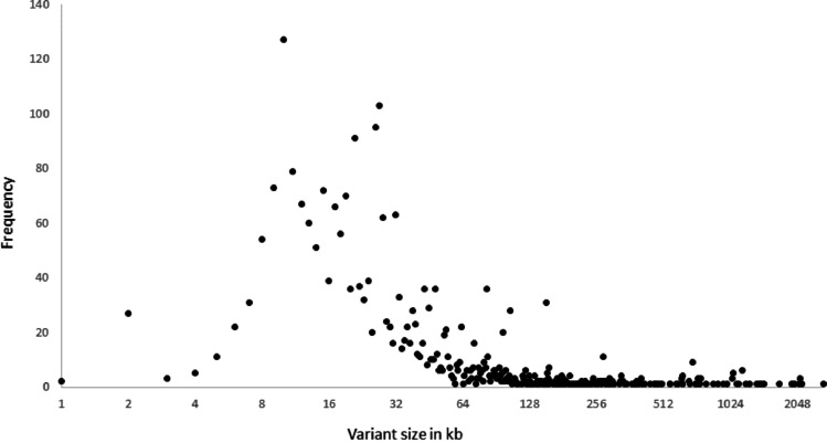
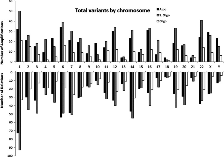
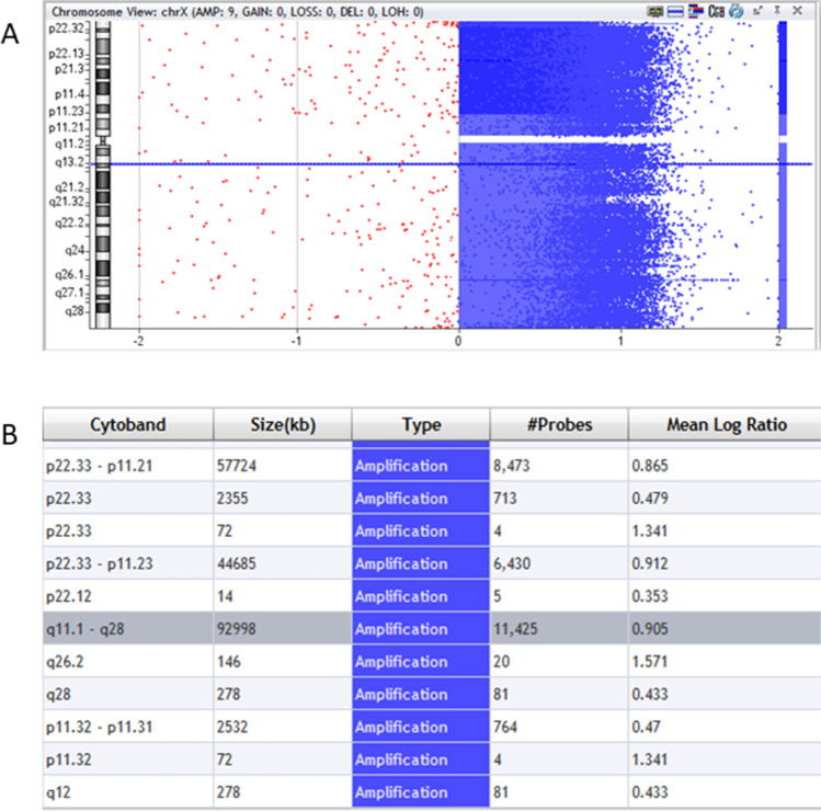
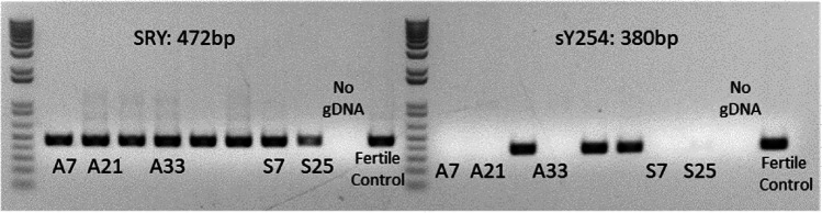
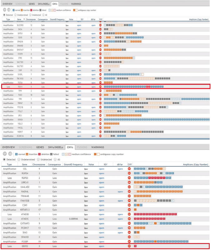
Results: Of the 2544 CNVs identified in individuals with SPGF, > 90% were small, ranging from 0.6 to 75 kb. Thirty, clinically relevant genomic aberrations, were detected in 28 patients (~ 29%). These included likely diagnostic CNVs in 3/41 NOA patients (~ 7%): 1 hemizygous, intragenic TEX11 deletion, 1 hemizygous DDX53 full gene deletion, and 1 homozygous, intragenic STK11 deletion. High-level mosaicism for X chromosome disomy (~ 10% 46,XY and ~ 90% 47,XXY) was also identified in 3 of 41 NOA patients who previously tested normal with conventional karyotyping. The remaining 24 CNVs detected were heterozygous, autosomal recessive carrier variants. Follow-up WES analysis confirmed 8 of 27 (30%) CNVs (X chromosome disomy excluded). WES analysis additionally identified 13 significant SNVs and/or indels in 9 patients (~ 9%) including X-linked AR, KAL1, and NR0B1 variants.

Conclusion: Using a combined genome-wide aCGH/WES approach, we identified pathogenic and likely pathogenic SNVs and CNVs in 15 patients (15%) with unexplained SPGF. This value equals the detection rate of conventional testing for aneuploidies and is considerably higher than the prevalence of Y chromosome microdeletions. Our results underscore the importance of comprehensive genomic analysis in emerging diagnostic testing of complex conditions like male infertility.

Keywords: Comparative genomic hybridization; Copy number variant; Single nucleotide variant; Spermatogenic failure; Whole-exome sequencing.


Conflict of interest statement

The authors declare no competing interests.

Figures

Fig. 1
Fig. 1
Patient variant size and frequency distribution. Total true variants (n = 2590) unique to individuals with spermatogenic failure (azoospermia n = 41, severe oligozoospermia n = 44, and oligozoospermia n = 17), detected by Agilent 400 K SurePrint G3 Human CGH array and calculated with Cytogenomics ADM-2 or ADM-2 Noisy algorithms, ranged in size from 0.6 kb-2.7 Mb, with > 95% of them less than 100 kb (X axis is shown in log2 scale)
Fig. 2
Fig. 2
Patient variant count and distribution per chromosome. Copy number variant detection by Agilent 400 K SurePrint G3 Human CGH array and quantification with Cytogenomics ADM-2 or ADM-2 Noisy algorithms showed no significant difference in average number of true CNVs per condition: azoospermia (n = 28), severe oligozoospermia (n = 28), oligozoospermia (n = 25). Total number of patient deletions (n = 1482) significantly outnumbered total number of patient amplifications (n = 1108) (p < 0.05). Normalization based on chromosome size showed no significant difference in chromosome distribution of copy number variants
Fig. 3
Fig. 3
Mosaic X chromosome amplification. A Agilent Cytogenomics chromosome view reveals amplification (blue) extending the entirety of the X chromosome. B Interval table. Highlighted row (gray) shows output for a 1.87-fold (2^0.905) amplification of a 93-Mb region of the X chromosome
Fig. 4
Fig. 4
Y Chromosome microdeletions detected by PCR. Agarose gel electrophoresis of amplified DNA shows the presence of SRY gene in eight patients and fertile control (left) and AZFc/sY254 (380 bp) deletions in samples A7, A21, A33, S7, and S25 (right)
Fig. 5
Fig. 5
WES confirmation of selected likely pathogenic CNVs. Output from SOPHiA DDM version 5.10.12 showing intragenic deletion of TEX11 (upper) and full deletion of DDX53 (lower) in two separate NOA patients

Similar articles

Cited by

References

    1. Krausz C, Riera-Escamilla A. Genetics of male infertility. Nat Rev Urol. 2018;15(6):369–384. doi: 10.1038/s41585-018-0003-3. - DOI - PubMed
    1. Matzuk MM, Lamb DJ. The biology of infertility: research advances and clinical challenges. Nat Med. 2008;14(11):1197–1213. doi: 10.1038/nm.f.1895. - DOI - PMC - PubMed
    1. Oud MS, et al. A systematic review and standardized clinical validity assessment of male infertility genes. Hum Reprod. 2019;34(5):932–941. doi: 10.1093/humrep/dez022. - DOI - PMC - PubMed
    1. Yatsenko AN, et al. Comprehensive 5-year study of cytogenetic aberrations in 668 infertile men. J Urol. 2010;183(4):1636–1642. doi: 10.1016/j.juro.2009.12.004. - DOI - PMC - PubMed
    1. Hardy JJ, et al. Variants in GCNA, X-linked germ-cell genome integrity gene, identified in men with primary spermatogenic failure. Hum Genet. 2021;140(8):1169–1182. doi: 10.1007/s00439-021-02287-y. - DOI - PMC - PubMed

Supplementary concepts