. 2022 Jul 18;17(7):e0271578.
doi: 10.1371/journal.pone.0271578. eCollection 2022.

Deletion of SM22α disrupts the structure and function of caveolae and T-tubules in cardiomyocytes, contributing to heart failure

Affiliations

Deletion of SM22α disrupts the structure and function of caveolae and T-tubules in cardiomyocytes, contributing to heart failure

Jun Wu et al. PLoS One. .

Abstract

Aims: Smooth muscle 22-alpha (SM22α) is an actin-binding protein that plays critical roles in mediating polymerization of actin filaments and stretch sensitivity of cytoskeleton in vascular smooth muscle cells (VSMCs). Multiple lines of evidence indicate the existence of SM22α in cardiomyocytes. Here, we investigated the effect of cardiac SM22α on the membrane architecture and functions of cardiomyocytes to pressure overload.

Methods: SM22α knock-out (KO) mice were utilized to assess the role of SM22α in the heart. Echocardiography was used to evaluate cardiac function, transverse aortic constriction (TAC) was used to induce heart failure, cell shortening properties were measured by IonOptix devices in intact cardiomyocytes, Ca2+ sensitivity of myofilaments was measured in permeabilized cardiomyocytes. Confocal microscopy, electron microscopy, western blotting, co-immunoprecipitation (co-IP), Real-Time Quantitative Reverse Transcription PCR (qRT-PCR) techniques were used to perform functional and structural analysis.

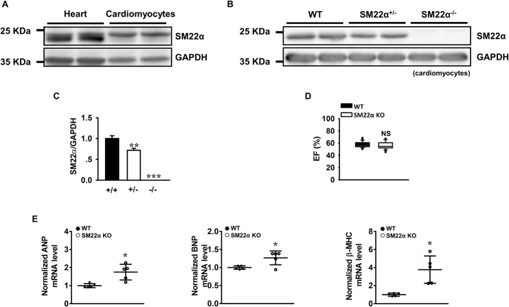
Results: SM22α ablation did not alter cardiac function at baseline, but mRNA levels of atrial natriuretic peptide (ANP), brain natriuretic peptide (BNP) and β-myosin heavy chain (β-MHC) were increased significantly compared with wild type (WT) controls. The membrane architecture was severely disrupted in SM22α KO cardiomyocytes, with disassembly and flattening of caveolae and disrupted T-tubules. Furthermore, SM22α was co-immunoprecipitated with caveolin-3 (Cav3), and the interaction between Cav3 and actin was significantly reduced in SM22α KO cells. SM22α KO cardiomyocytes displayed asynchronized SR Ca2+ release, significantly increased Ca2+ spark frequency. Additionally, the kinetics of sarcomere shortening was abnormal, accompanied with increased sensitivity and reduced maximum response of myofilaments to Ca2+ in SM22α KO cardiomyocytes. SM22α KO mice were more prone to heart failure after TAC.

Conclusions: Our findings identified that SM22α may be required for the architecture and function of caveolae and T-tubules in cardiomyocytes.


Conflict of interest statement

The authors have declared that no competing interests exist.

Figures

Fig 1
Fig 1. The cardiac function is reduced in SM22α KO mice.
(A) and (B) Representative Western blot of the expression of SM22α protein in heart tissue and cardiomyocytes of WT, SM22α KO (SM22α-/-) and heterozygous (SM22α+/-) mice. (C) Bar graphs showing the expression of SM22α protein in WT (+/+), heterozygous (+/-) and KO (-/-) mice (n = 6 per group). (D) Echocardiographic assessment of ejection fraction (EF) (n = 40 per group). (E) RT-PCR analysis of cardiac fetal genes and hypertrophic markers ANP, BNP, β-MHC (n = 5 per group). All data are represented as mean ± SEM from 3 independent experiments. *P< 0.05, **P< 0.01, ***P< 0.001 vs. WT control.
Fig 2
Fig 2. Disassembly and flattening of caveolae occur in the cardiomyocytes from SM22α KO mice.
(A, B) Low-magnification electron micrographs of caveolae in the cardiomyocytes from WT (A) and SM22α KO (B) mice (arrowheads denote caveolae), Scale bars = 500 nm. (C) Bar graphs showing decreased caveolae density in the cardiomyocyte of SM22α KO mice (WT: 22 micrographs from 3 mice; KO: 27 micrographs from 3 mice). (D, E) Zoom-in images of representative caveolae. Yellow dashed-lines: highlighting the shape of representative caveolae. Scale bars = 100 nm. (F) Bar graphs showing increased opening width of caveolae in cardiomyocytes of SM22α KO mice (n = 67 caveolae from 3 WT mice and n = 77 caveolae from 3 KO mice). (G, H) Bar graphs showing no significant changes of caveolae maximal width (G) and depth (H) in cardiomyocytes of SM22α KO mice (n = 67 caveolae from 3 WT mice and n = 77 caveolae from 3 KO mice). Data are presented as the mean ± SEM. **P< 0.01, ***P< 0.001 vs. WT control.
Fig 3
Fig 3. The interaction between actin and Cav3 is decreased in SM22α KO cardiomyocyte.
(A) Western blot for the expression of Cav3. (B, C) Co-immunoprecipitation (co-IP) analysis for the interaction between SM22α, Cav3 and α-actin. Input lanes correspond to the original extracts used for the co-IP assays. IgG was used as negative control. (D) Bar graphs showed the quantification of α-actin-Cav3 interaction. WCL: whole cell lysate. Data are presented as the mean ± SEM from 3 independent experiments (n = 3). *P< 0.05 vs. WT control.
Fig 4
Fig 4. T-tubules are disrupted in SM22α KO cardiomyocytes.
(A) Confocal fluorescence images of representative di-8-ANEPPS-stained single cardiomyocytes isolated from WT and SM22α KO mice (upper). Respective high-resolution views and of areas within the red rectangles (middle). Corresponding Fast Fourier transformed images of T-tubule images (bottom). Scale bar = 10 μm. (B) Representative power vs. spatial frequency along the x axis computed using Fourier analysis. (C) Bar graph showing TT score vs. WT control (n = 20 cells from 3 hearts in each group). (D) qRT-PCR analysis of JPH2 mRNA levels (WT: n = 9; KO: n = 8). (E) Western blot of JPH2 protein level in cardiomyocytes of WT and SM22α KO mice. (F) Bar graph showed the mean ± SEM from 3 independent experiments (n = 4 per group). (G) Co-IP analysis for the interaction of SM22α with JPH2. *P< 0.05, ***P< 0.001 vs. WT control.
Fig 5
Fig 5. The disruption of SM22α is associated with abnormal SR Ca2+ handling in the cardiomyocytes.
(A) Confocal microscopy line scans revealing increased number of Ca2+ sparks in SM22α KO mice. (B) Bar graph showing frequency of spontaneous Ca2+ sparks (n = 15 cells from 3 WT mice and 16 cells from 3 SM22α KO mice). (C) Representative tracings of Ca2+ transient amplitudes when paced at 1 Hz and total SR Ca2+ content as measured by the amplitude of caffeine-induced Ca2+ transient. (D) Bar graphs showing average SR Ca2+ content (n = 10 cells from 3 WT mice and 8 cells from 3 SM22α KO mice). (E) Representative confocal line scan images showing desynchronized Ca2+ transient in SM22α KO mice. (F) Bar graphs showing the average Ca2+ transient amplitude (n = 23 cells from 3 WT mice and 20 cells from 3 SM22α KO mice). (G) Bar graphs showing the Ca2+ decay constant (τ) (n = 23 cells from 3 WT mice and 20 cells from 3 SM22α KO mice). (H) Bar graphs showing the time to peak (TtP) (n = 23 cells from 3 WT mice and 20 cells from 3 SM22α KO mice). Data are presented as the mean ± SEM. *P< 0.05 vs. WT control.
Fig 6
Fig 6. SM22α KO cardiomyocytes display altered kinetics of cell shortening.
(A) Representative tracings showed altered kinetics of cell shortening in SM22α KO cardiomyocytes. (B) The amplitude of contraction of the cardiomyocytes. (C-E) Bar graphs showing decreased TtP (C), increased Dep v (D) and reduced Dep v t (E) in SM22α KO mice (n = 72 cells from 6 WT mice; n = 31 cells from 4 SM22α KO mice). *P< 0.05, ***P< 0.001 vs. WT control. (F) Perfused cells with 0, 200, 300, 400, 500 nmol/L Ca2+ buffer to record cell shortening in saponin permeabilized myocytes. Continuous lines showed Hill fits with Hill coefficients (nH) = -6.22 and concentration for 50% of maximal effect (EC50) = 515 nmol/L for WT cells, and nH = -4.99, EC50 = 288 nmol/L for the KO cells (n = 9 or 10 from 3 mice in each group). Ca2+ sensitivity of myofilament was increased (right), but maximum response of myofilament was decreased in SM22α KO cells (left). Data are presented as the mean ± SEM. *P< 0.5, ***P< 0.001 vs. WT control.
Fig 7
Fig 7. The disruption of SM22α accelerates the development of heart failure induced by TAC.
(A) Western blot of SM22α in cardiomyocytes of WT mice sham and TAC hearts. (B) Bar graph showed increased level of SM22α protein post TAC (n = 5 in each group). (C) qRT-PCR analysis of SM22α mRNA (WT: n = 21; KO: n = 12). (D) Echocardiographic assessment of heart function using M-mode images of left ventricle. (E) Quantitative evaluation of systolic function in LV EF of 0, 2, 4 and 8 weeks post TAC (n = 35–40 per group). Data are presented as the mean ± SEM. *P< 0.5, ***P < 0.001 vs. WT control for the same week, ###P< 0.001 vs. WT baseline, +++P< 0.001 vs. SM22α KO baseline. (F) Kaplan-Meier curve revealing increased mortality in SM22α KO mice compared with WT mice after TAC (WT: n = 20; KO: n = 26). (G) Whole mount of representative hearts from WT and SM22α KO sham or 4 weeks post TAC. (H, I) Heart weight to body weight ratio (HW/BW) (H) and lung weight to body weight ratio (LW/BW) (I) for WT and SM22α KO mice 2 and 4 weeks post TAC or sham surgery (n = 6–10 per group). Data are represented as mean ± SEM. *P< 0.05, **P< 0.01, ***P< 0.001.

Similar articles

References

    1. Shapland C, Hsuan JJ, Totty NF, Lawson D. Purification and properties of transgelin: a transformation and shape change sensitive actin-gelling protein. J Cell Biol. 1993;121(5):1065–1073. doi: 10.1083/jcb.121.5.1065 - DOI - PMC - PubMed
    1. Li L, Liu Z, Mercer B, Overbeek P, Olson EN. Evidence for serum response factor-mediated regulatory networks governing SM22alpha transcription in smooth skeletal and cardiac muscle cells. 1997. J;187(2):311–321. doi: 10.1006/dbio.1997.8621 - DOI - PubMed
    1. Lees-Miller JP, Heeley DH, Smillie LB. An abundant and novel protein of 22 kDa (SM22) is widely distributed in smooth muscles. Purification from bovine aorta. Biochem J. 1987;244(3):705–709. doi: 10.1042/bj2440705 - DOI - PMC - PubMed
    1. Lees-Miller JP, Heeley DH, Smillie LB, Kay CM. Isolation and characterization of an abundant and novel 22-kDa protein (SM22) from chicken gizzard smooth muscle. J Biol Chem. 1987;262(7):2988–2993.. - PubMed
    1. Zeidan A, Swärd K, Nordström I, Ekblad E, Zhang JCL, Parmacek MS, et al.. Ablation of SM22alpha decreases contractility and actin contents of mouse vascular smooth muscle. FEBS Lett. 2004;562(1–3):141–146. doi: 10.1016/S0014-5793(04)00220-0 - DOI - PubMed

Publication types