. 2022 Jul 18;16(7):e0010618.
doi: 10.1371/journal.pntd.0010618. eCollection 2022 Jul.

2-Deoxy-D-glucose and combined 2-Deoxy-D-glucose/albendazole exhibit therapeutic efficacy against Echinococcus granulosus protoscoleces and experimental alveolar echinococcosis

Affiliations

2-Deoxy-D-glucose and combined 2-Deoxy-D-glucose/albendazole exhibit therapeutic efficacy against Echinococcus granulosus protoscoleces and experimental alveolar echinococcosis

Qi Xin et al. PLoS Negl Trop Dis. .

Abstract

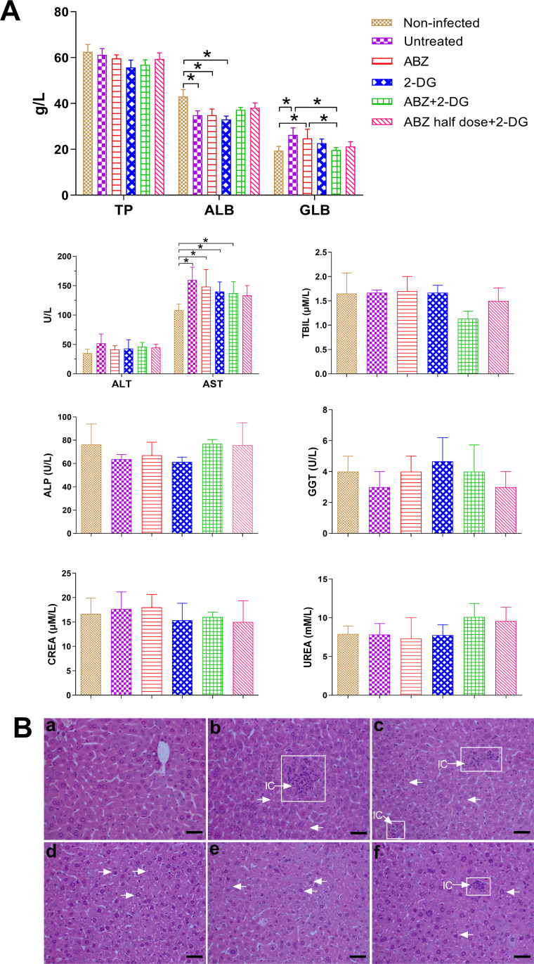
2-Deoxy-D-glucose (2-DG) is a glucose analog used as a promising anticancer agent. It exerts its effects by inhibiting the glycolytic energy metabolism to deplete cells of energy. The larval stage of Echinococcus relies on glycolysis for energy production. Therefore, in this study, we investigated the in vitro and in vivo efficacy of 2-DG against the larval stage of Echinococcus granulosus and E. multilocularis. 2-DG exhibited significant time- and dose-dependent effects against in vitro cultured E. granulosus protoscoleces and E. multilocularis metacestodes. A daily oral administration of 500 mg/kg 2-DG in E. multilocularis-infected mice effectively reduced the weight of metacestodes. Notably, the combination treatment, either 2-DG (500 mg/kg/day) + albendazole (ABZ) (200 mg/kg/day) or 2-DG (500 mg/kg/day) + half-dose of ABZ (100 mg/kg/day), exhibited a potent therapeutic effect against E. multilocularis, significantly promoting the reduction of metacestodes weight compared with the administration of 2-DG or ABZ alone. Furthermore, the combination significantly promoted apoptosis of the cells of metacestodes and inhibited glycolysis in metacestodes, compared with the administration of 2-DG or ABZ alone. In conclusion, 2-DG exerts an effective activity against the larval stage of Echinococcus. Thus, it may be a promising anti-Echinococcus drug, and its combination with ABZ may provide a new strategy for the treatment of echinococcosis in humans.


Conflict of interest statement

The authors have declared that no competing interests exist.

Figures

Fig 1
Fig 1. In vitro effect of 2-DG against E. granulosus protoscoleces.
(A) Viability of protoscoleces incubated for 7 days with 40 μM NTZ or serial concentrations of 2-DG (10, 20, 40, 80, 160, and 320 μM). Protoscoleces incubated in a culture medium containing 0.2% DMSO were used as a control. (B) Scanning electron microscopy of protoscoleces after 5 days of incubation. (a) Invaginated control protoscoleces; (b) Evaginated control protoscoleces (rr, rostelar region; s, suckers; sr, soma region); (c) Protoscoleces incubated with 40 μM 2-DG. Protoscoleces showed marked contraction, shedding of microtriches and loss of hooks (arrow).
Fig 2
Fig 2. In vitro effects of 2-DG on E. multilocularis metacestodes.
(A) E. multilocularis AP (EmAP) activities in the medium supernatant after 36 and 120 hours of treatment with 40 μM NTZ or various 2-DG concentrations (10, 20, 40, 80, 160, and 320 μM). Vesicles incubated in the culture medium containing DMSO served as controls. *p < 0.05 vs. DMSO control. (B) Microscopical damage to metacestodes treated with 40 μM NTZ or 40 μM 2-DG for 36 and 120 hours. Control vesicles exhibited no morphological changes, while drug-treated vesicles showed increased collapse and wrinkled cyst walls. LL, laminated layer; GL, germinal layer. Scale bar, 200 μm.
Fig 3
Fig 3. In vivo effect of 2-DG and its combination with ABZ in E. multilocularis–infected mice.
(A) The weights and gross morphology of the metacestodes in untreated mice and mice treated with drugs for 6 weeks. *p < 0.05 vs. untreated mice. (B) Histopathological examination of E. multilocularis metacestodes from different treatment groups. a, mice infected with E. multilocularis; b, c, d, and e, infected mice treated with ABZ (200 mg/kg/day), 2-DG (500 mg/kg/day), ABZ + 2-DG (200 mg/kg/day + 500 mg/kg/day), and ABZ half-dose + 2-DG (100 mg/kg/day + 500 mg/kg/day), respectively. LL, laminated layer; GL, germinal layer; IC, inflammatory cell; FT, fibrous tissue. Scale bar, 50 μm.
Fig 4
Fig 4. Cytokine profiles and histopathological examination.
Serum concentrations of IFN-γ and IL-10 (A) were measured, and spleen tissues were observed (H&E staining) (B) from mice in different treatment groups. a, non-infected mice; b, mice infected with E. multilocularis metacestodes; c, d, e, and f, infected mice treated with ABZ, 2-DG, ABZ + 2-DG, and ABZ half-dose + 2-DG, respectively. RePu, the red pulp region in the spleen; WhPu, the white pulp region (lymphoid tissue containing lymphocyte production centers) in the spleen. *p < 0.05 vs. untreated mice, ▲p < 0.05 vs. non-infected mice. Scale bar, 50 μm.
Fig 5
Fig 5. Ultrastructural alterations of metacestodes in different treatment groups.
(A) Scanning electron microscopy (SEM) and transmission electron microscopy (TEM) (B and C) show metacestodes from untreated mice. The metacestode has a characteristic structure with an intact germinal layer surrounded by an acellular laminated layer. The arrows indicate microtriches originating from the tegument and protruding into the laminated layer. Note the large number and the length of microtriches. (D) SEM and TEM (E and F) show metacestodes from ABZ-treated mice. Note the complete absence of microtriches at the tegument and regression of the germinal layers. (G) SEM and TEM (H and I) show metacestodes from 2-DG–treated mice. Note the profound shortening and reduced number of the microtriches (marked by a white box and arrows) and a less dense appearance of the germinal layer tissue. Moreover, the characteristic features of apoptosis are visible in undifferentiated cells (marked by a red box). (J), (K), (L) and (M), (N), and (O) show metacestodes from mice treated with ABZ + 2-DG and ABZ half-dose + 2-DG. The metacestodes exhibited complete breakdown of the structural integrity of the vesicles; there was a complete loss of microtriches, and only residual laminated layers lacking any cellular parasites components were found in many areas. LL, laminated layer; GL, germinal layer; Teg, tegument; Uc, undifferentiated cells; gly, glycogen storage cells; vac, vacuolization of the tissue; nu, cell nucleus; ld, lipid drops.
Fig 6
Fig 6. Serum biochemical analysis and histopathological examination in mice treated with ABZ, 2-DG, ABZ + 2-DG, or ABZ half-dose + 2-DG for 6 weeks.
(A) The levels of total protein (TP), albumin (ALB), globulin (GLB), alanine aminotransferase (ALT), aspartate aminotransferase (AST), total bilirubin (TBIL), alkaline phosphatase (ALP), glutamyltranspeptidase (GGT), urea, and creatinine (CREA) in mice serum. *p < 0.05. (B) H&E images of liver tissues. Non-infected tissue (a), E. multilocularis-infected tissue (b), ABZ-treated tissue (c), 2-DG-treated tissue (d), ABZ + 2-DG–treated tissue (e), and ABZ half-dose + 2-DG–treated tissue (f) were visualized with a microscope. Focal necrosis is marked by a white box, and the white arrows indicate hepatocyte edema. IC, inflammatory cell. Scale bar, 50 μm.
Fig 7
Fig 7. Analysis of apoptosis in E. multilocularis metacestodes.
The TUNEL staining was performed in metacestodes from untreated mice, mice treated with ABZ (200 mg/kg/day), 2-DG (500 mg/kg/day), ABZ + 2-DG (200 mg/kg/day + 500 mg/kg/day), or ABZ half-dose + 2-DG (100 mg/kg/day + 500 mg/kg/day). Apoptosis was evaluated by the integrated density of green fluorescence in metacestodes using ImageJ software. *p < 0.05 vs. untreated mice.
Fig 8
Fig 8. Effects of ABZ, 2-DG, ABZ + 2-DG, or ABZ half-dose + 2-DG on glucose metabolism of E. multilocularis metacestodes.
Various concentrations of ABZ, 2-DG, or combined ABZ/2-DG were preincubated with 200 μg metacestodes slices for 30 minutes at 37°C, and then the slices were incubated in the presence of 6 mM glucose. (A) Glucose consumption was determined for the times indicated. *p < 0.05, 2-DG 200 mM–treated group vs. control group, ABZ 320 μM, ABZ 80 μM, or ABZ 20 μM–treated group. **p < 0.05, ABZ 320 μM + 2-DG 200 mM vs. control group, ABZ 320 μM, ABZ 80 μM, or ABZ 20 μM–treated group. #p < 0.05, ABZ 160 μM + 2-DG 200 mM vs. control group, ABZ 320 μM, ABZ 80 μM, or ABZ 20 μM–treated group. ▲p < 0.05, 2-DG 100 mM–treated group vs. control group or ABZ 80 μM–treated group. (B) Lactate production in culture medium for the times indicated. *p < 0.05 vs. control group.

Similar articles

Cited by

References

    1. Cadavid Restrepo AM, Yang YR, McManus DP, Gray DJ, Giraudoux P, Barnes TS, et al.. The landscape epidemiology of echinococcoses. Infect Dis Poverty. 2016; 5: 13. doi: 10.1186/s40249-016-0109-x - DOI - PMC - PubMed
    1. Deplazes P, Rinaldi L, Alvarez Rojas CA, Torgerson PR, Harandi MF, Romig T, et al.. Global distribution of alveolar and cystic echinococcosis. Adv Parasitol. 2017; 95: 315–493. doi: 10.1016/bs.apar.2016.11.001 - DOI - PubMed
    1. Kern P, Menezes da Silva A, Akhan O, Müllhaupt B, Vizcaychipi KA, Budke C, et al.. The echinococcoses: diagnosis, clinical management and burden of disease. Adv Parasitol. 2017; 96: 259–369. doi: 10.1016/bs.apar.2016.09.006 - DOI - PubMed
    1. Wen H, Vuitton L, Tuxun T, Li J, Vuitton DA, Zhang W, et al.. Echinococcosis: Advances in the 21st Century. Clin Microbiol Rev. 2019; 32(2): e00075–18. doi: 10.1128/CMR.00075-18 - DOI - PMC - PubMed
    1. Hemphill A, Stadelmann B, Rufener R, Spiliotis M, Boubaker G, Müller J, et al.. Treatment of echinococcosis: albendazole and mebendazole—what else? Parasite. 2014; 21: 70. doi: 10.1051/parasite/2014073 - DOI - PMC - PubMed

Publication types

Supplementary concepts