. 2023 Mar 21;33(7):3401-3420.
doi: 10.1093/cercor/bhac280.

Estrogens rapidly shape synaptic and intrinsic properties to regulate the temporal precision of songbird auditory neurons

Affiliations

Estrogens rapidly shape synaptic and intrinsic properties to regulate the temporal precision of songbird auditory neurons

Garrett B Scarpa et al. Cereb Cortex. .

Abstract

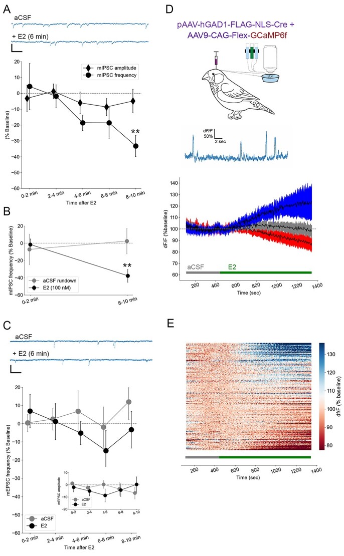
Sensory neurons parse millisecond-variant sound streams like birdsong and speech with exquisite precision. The auditory pallial cortex of vocal learners like humans and songbirds contains an unconventional neuromodulatory system: neuronal expression of the estrogen synthesis enzyme aromatase. Local forebrain neuroestrogens fluctuate when songbirds hear a song, and subsequently modulate bursting, gain, and temporal coding properties of auditory neurons. However, the way neuroestrogens shape intrinsic and synaptic properties of sensory neurons remains unknown. Here, using a combination of whole-cell patch clamp electrophysiology and calcium imaging, we investigate estrogenic neuromodulation of auditory neurons in a region resembling mammalian auditory association cortex. We found that estradiol rapidly enhances the temporal precision of neuronal firing via a membrane-bound G-protein coupled receptor and that estradiol rapidly suppresses inhibitory synaptic currents while sparing excitation. Notably, the rapid suppression of intrinsic excitability by estradiol was predicted by membrane input resistance and was observed in both males and females. These findings were corroborated by analysis of in vivo electrophysiology recordings, in which local estrogen synthesis blockade caused acute disruption of the temporal correlation of song-evoked firing patterns. Therefore, on a modulatory timescale, neuroestrogens alter intrinsic cellular properties and inhibitory neurotransmitter release to regulate the temporal precision of higher-order sensory neurons.

Keywords: aromatase; cortex; estradiol; learning; neuromodulation; zebra finch.


Figures

Fig. 1
Fig. 1
Electrical phenotype classifications during current-clamp recordings segregate NCM neurons by rheobase and IR. A) Representative voltage traces illustrating classification differences between transient/phasic neurons versus tonic neurons in NCM. Rheobase is plotted in black, all other voltage traces plotted in gray; only a subset of voltage traces and current-step injections are plotted for clarity. Plotted current steps are 50 pA. Scale bar = 10 mV; 100 ms. Resting membrane potential (B, RMP), AP peak (C), AP half-width (D, AP HW), and AP repolarization tau (E) were not different between tonic (N = 19 cells) versus transient/phasic (N = 11 cells) cell type categories in response to current stimulation. By contrast, rheobase (F) and IR (G) were each significantly different between the 2 classification schemes. *P < 0.05.
Fig. 2
Fig. 2
Only NCM neurons with low IR respond to bath-applied 17-beta-estradiol (E2). A, B) Differential change in passive membrane conductance (IR) among high-IR (N = 8) versus low-IR (N = 11) neurons in NCM in response to bath application of 50 nM E2. In panel A, numbers in parentheses denote time in minutes following E2 application. C) Top: Representative waveforms during recordings of spontaneous activity in low-IR versus high-IR neurons. Scale bars = 10 mV, 10 ms. Bottom: Histogram of IR from the population of recorded cells, with a cluster split at 400 MOhm (colors denote one-dimensional k-means algorithm via Scikit-learn Python library; (Pedregosa et al. 2011). D) Properties of high- versus low-IR NCM neurons in terms of sag index (D), resting membrane potential (E), AP half-width (F), and AP repolarization tau (G). *P < 0.05.
Fig. 3
Fig. 3
Estradiol (E2) rapidly changes intrinsic excitability and temporal precision in NCM neurons with low IR. A) Representative current-clamp traces for a low-IR (left) versus high-IR (right) neurons in NCM. Note the changes in active and passive membrane properties in response to 50 nM E2 in low-IR neurons exclusively, including spiking activity (peak firing) as well as IR (inset numbers) for each cell. B) The peak firing (maximum number of spikes evoked per series of current steps; expressed as a % change from the 10 min baseline period) of low-IR neurons (N = 11) drops significantly 8–10 min following bath application of 50 nM E2, while peak firing is not significantly altered in high-IR neurons (N = 8). Numbers on x-axis denote time in minutes relative to E2 application. Inset: Similar magnitude decrease in peak firing of low-IR neurons observed in males and females. C) F/I relationships during baseline (aCSF) and following 8–10 min of 50 nM E2 (+E2) for low-IR (left) versus high-IR (right) neurons. The firing rate (FR) of low-IR neurons was suppressed by 50 nM E2 at all depolarizing current steps from 40 to 80 pA, while no changes on FR were observed in high-IR neurons. D, E) The temporal precision of the first AP evoked at rheobase in low-IR neurons was also enhanced by 50 nm E2, reflected in a quickening of first-AP latency (D) and reduction in first-AP latency jitter (E) in low- but not high-IR neurons. Numbers in parentheses denote time in minutes following E2 application. F) Rundown control experiments in low-IR neurons showing no change in either IR (top) or peak firing (bottom). ***P < 0.001, **P < 0.005.
Fig. 4
Fig. 4
Aromatase expression is not restricted to high- or low-IR neurons. A) Recorded low-IR neurons which did express aromatase (top) and those which did not (bottom) despite surrounding aromatase positive cells. B) Recorded high-IR neurons which did express aromatase (top) and those which did not (bottom) despite surrounding aromatase positive cells. For each row, green (left; 488 nm) is internal solution neurobiotin filled during current-clamp recordings, while red (middle; 594 nm) is antibody-labeled aromatase (merge at right). Scale bars = 10 μm.
Fig. 5
Fig. 5
Estradiol rapidly suppresses miniature inhibitory postsynaptic currents. Bath-applied E2 at 50 nM (A; N = 13) and 100 nM (B; N = 4) causes a 30%–40% decrease in the frequency of miniature inhibitory postsynaptic currents (mIPSCs) but not their amplitude (A) and no change among rundown controls (B; N = 6). **P < 0.01 for within-cell differences from baseline. At top of (A) are plotted representative raw current traces of mIPSCs from NCM neurons (scale bar = 20 pA; 25 ms). By contrast, there were no changes in frequency or amplitude (inset) of miniature excitatory post-synaptic currents (mEPSCs) for either aCSF controls (N = 5) or 50 nM E2 (C; N = 13). At top of (C) are plotted representative raw current traces of mEPSCs from NCM neurons (scale bar = 20 pA; 25 ms). (D) Calcium imaging (% of baseline) reflecting spontaneous cellular and network excitability revealed 3 distinct responses to 50 nM E2 among GAD1-GCaMP6-expressing inhibitory neurons (n = 288 ROIs): 29% of units displayed a ~30% increase in activity (blue), 20% demonstrated a ~15% decrease in activity (red), and 51% remained stable (gray). Data were binned at 34 s, and criterion for an increase or decrease in activity was a ±10% change in 3 out of 4 consecutive bins during the treatment period. Top, schematic showing viral injection and ex vivo calcium imaging, and a line-scan imaging session with large, spike-like spontaneous intracellular Ca2+ events (D). Same results as shown in (D) are replotted with all R0Is and sorted in a heat map (E).
Fig. 6
Fig. 6
Rapid estradiol effects require GPER1 activation, and suppressing in vivo brain estrogen synthesis disrupts NCM temporal firing correlations. A) Antagonist experiments with the GPER1 blocker G36 (N = 5) indicate that acute E2-dependent changes in peak firing (top) and IR (bottom) in low-IR neurons are dependent on GPER1. At top are raw voltage trace overlays of representative cells at 5 and 20 min following bath application of G36 alone or G36 in combination with E2. The peak firing of these neurons to +140 pA current injections (top) and hyperpolarization to −200 pA current injections (bottom) are shown here. Scale bar = 250 ms; 15 mV. B) Retrodialysis of fadrozole (FAD), coupled with playback of conspecific birdsong, resulted in a drop in trial-by-trial correlation of song-evoked firing, in vivo. Top, representative raster plot (top), peristimulus time histogram (middle), and song oscillogram (bottom). Bottom mean ± SEM of trial-by-trial correlation for aCSF versus 100 μM FAD retrodialysis (N = 12 cells). Dashed line is baseline trial-by-trial correlation of neurons in the absence of song playback. ***P < 0.005 for within-unit difference from aCSF. At top is 100 overlays of isolated waveforms from a single extracellular unit in NCM.

Similar articles

Cited by

References

    1. Acharya KD, Veney SL. Characterization of the G-protein-coupled membrane-bound estrogen receptor GPR30 in the zebra finch brain reveals a sex difference in gene and protein expression. Dev Neurobiol. 2012:72(11):1433–1446. - PubMed
    1. Alcami P. Electrical synapses enhance and accelerate interneuron recruitment in response to coincident and sequential excitation. Front Cell Neurosci. 2018:12:156. - PMC - PubMed
    1. Azcoitia I, Yague JG, Garcia-Segura LM. Estradiol synthesis within the human brain. Neuroscience. 2011:191:139–147. - PubMed
    1. Bailey DJ, Saldanha CJ. The importance of neural aromatization in the acquisition, recall, and integration of song and spatial memories in passerines. Horm Behav. 2015:74:116–124. - PMC - PubMed
    1. Balthazart J, Ball G. Is brain estradiol a hormone or a neurotransmitter? Trends Neurosci. 2006:29(5):241–249. - PubMed

Publication types