Review
. 2022 Oct 6:38:395-418.
doi: 10.1146/annurev-cellbio-120420-114855. Epub 2022 Jul 18.

Tissue Homeostasis and Non-Homeostasis: From Cell Life Cycles to Organ States

Affiliations
Review

Tissue Homeostasis and Non-Homeostasis: From Cell Life Cycles to Organ States

Lucy Erin O'Brien. Annu Rev Cell Dev Biol. .

Abstract

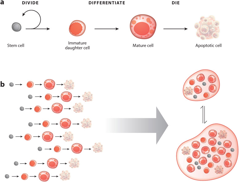
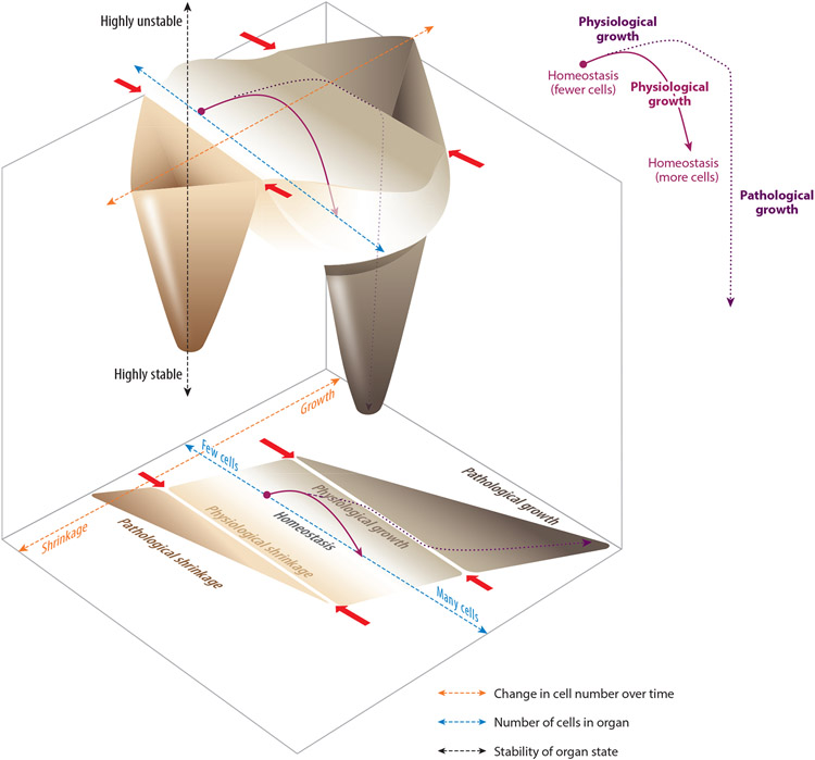
Although tissue homeostasis-the steady state-implies stability, our organs are in a state of continual, large-scale cellular flux. This flux underpins an organ's ability to homeostatically renew, to non-homeostatically resize upon altered functional demand, and to return to homeostasis after resizing or injury-in other words, to be dynamic. Here, I examine the basic unit of organ-scale cell dynamics: the cellular life cycle of birth, differentiation, and death. Focusing on epithelial organs, I discuss how spatial patterns and temporal kinetics of life cycle stages depend upon lineage organization and tissue architecture. I review how signaling between stages coordinates life cycle dynamics to enforce homeostasis, and I highlight how particular stages are transiently unbalanced to drive organ resizing or repair. Finally, I offer that considering organs as a collective of not cells but rather cell life cycles provides a powerful vantage for deciphering homeostatic and non-homeostatic tissue states.

Keywords: adaptation; differentiation; extrusion; feedback; stem cells; tissue homeostasis.


Figures

Figure 1
Figure 1
The cell life cycle, which encompasses (a) birth (via division of a stem cell), terminal differentiation, mature physiological function, and apoptotic death, can be regarded as (b) the basic unit of organ-scale cell dynamics.
Figure 2
Figure 2
Hypothetical organ state landscapes in two and three dimensions, based on the parameter of cell number. (Bottom) The two-dimensional landscape shows total cell number (blue dashed axis) relative to rate of change in total cell number (orange dashed axis). Within this parameter space, approximate regions are indicated for organ states of homeostasis, physiological growth, pathological growth, physiological shrinkage, and pathological shrinkage. (Top) In the corresponding three-dimensional landscape, homeostasis, pathological growth, and pathological shrinkage are drawn as basins, with the depth of the basin indicating a state’s relative stability (black dashed axis). Physiological growth and physiological shrinkage are represented by the slopes above the floor of the homeostasis basin. Finally, the boundaries between physiological and pathological growth or shrinkage are represented by the ridge tops separating the basins (red arrows). Two hypothetical examples of organ dynamics are illustrated by the purple lines (shown within the landscapes and also isolated to the right). The solid purple line shows a physiological growth trajectory. The organ is initially homeostatic but contains relatively few cells (perhaps, for instance, because it is experiencing low functional demand). In response to environmental change (for instance, an increase in functional demand), the organ undergoes physiological growth and acquires additional cells until it reaches an appropriate, larger size. At this point, the organ ceases growth and returns to homeostasis, but at its new, larger size. By contrast, the dotted purple line shows an organ in which growth becomes pathological. The organ acquires a huge excess of additional cells, crosses the boundary between physiological and pathological growth, and ultimately becomes deeply stuck in a maladaptive state of overgrowth.

References

    1. Akhtar N, Li W, Mironov A, Streuli CH. 2016. Rac1 controls both the secretory function of the mammary gland and its remodeling for successive gestations. Dev. Cell 38(5):522–35 - PMC - PubMed
    1. Aldewachi HS, Wright NA, Appleton DR, Watson AJ. 1975. The effect of starvation and refeeding on cell population kinetics in the rat small bowel mucosa. J. Anat 119(Part 1):105–21 - PMC - PubMed
    1. Altmann GG. 1972. Influence of starvation and refeeding on mucosal size and epithelial renewal in the rat small intestine. Am. J. Anat 133(4):391–400 - PubMed
    1. Amcheslavsky A, Ito N, Jiang J, Ip YT. 2011. Tuberous sclerosis complex and Myc coordinate the growth and division of Drosophila intestinal stem cells. J. Cell Biol 193(4):695–710 - PMC - PubMed
    1. Amcheslavsky A, Lindblad JL, Bergmann A. 2020. Transiently “undead” enterocytes mediate homeostatic tissue turnover in the adult Drosophila midgut. Cell Rep. 33(8):108408. - PMC - PubMed

Publication types

LinkOut - more resources