Review
. 2022 Oct 5;55(5):364-377.
doi: 10.5090/jcs.22.030. Epub 2022 Jul 20.

Surgical and Electrical Anatomy of the Inter-Nodal and Intra-Atrial Conduction System in the Heart

Affiliations
Review

Surgical and Electrical Anatomy of the Inter-Nodal and Intra-Atrial Conduction System in the Heart

Jeong-Wook Seo et al. J Chest Surg. .

Abstract

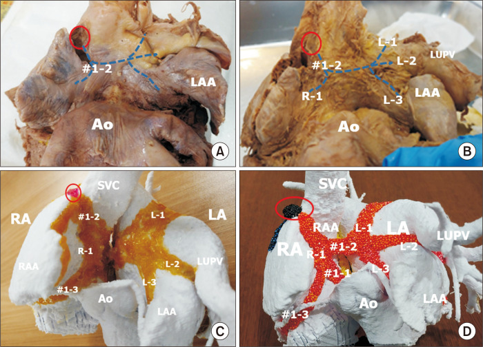
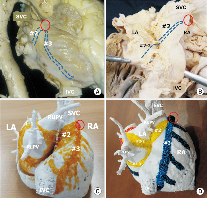
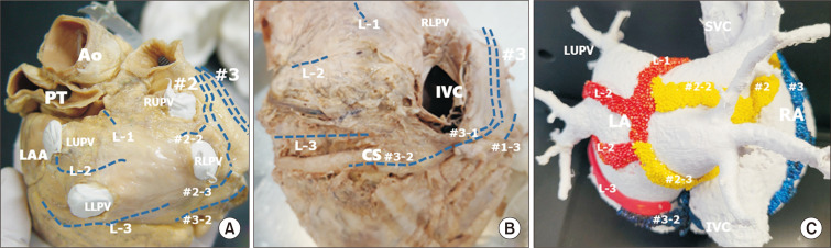
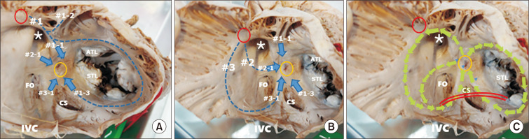
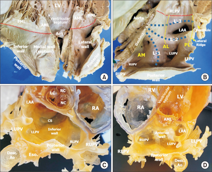
An anatomical understanding of the atrial myocardium is crucial for surgeons and interventionists who treat atrial arrhythmias. We reviewed the anatomy of the inter-nodal and intra-atrial conduction systems. The anterior inter-nodal route (#1) arises from the sinus node and runs through the ventral wall of the atrial chambers. The major branch of route #1 approaches the atrioventricular node from the anterior aspect. Other branches of route #1 are Bachmann's bundle and a vestibular branch around the tricuspid valve. The middle inter-nodal route (#2) begins with a broad span of fibers at the sinus venarum and extends to the superior limbus of the oval fossa. The major branch of route #2 joins with the branch of route #1 at the anterior part of the atrioventricular node. The posterior inter-nodal route (#3) is at the terminal crest and gives rise to many branches at the pectinate muscles of the right atrium and then approaches the posterior atrioventricular node after joining with the vestibular branch of route #1. The branches of the left part of Bachmann's bundle and the branches of the second inter-nodal route form a thin myocardial network at the posterior wall of the left atrium. These anatomical structures could be categorized into major routes and side branches. There are 9 or more anatomical circles in the atrial chambers that could be structural sites for macro re-entry. The implications of normal and abnormal structures of the myocardium for the pathogenesis and treatment of atrial arrhythmias are discussed.

Keywords: Atrial fibrillation; Atrial flutter; Bachmann’s bundle; Cardiac arrhythmia; Cardiac conduction system; Maze procedure; Radiofrequency ablation.


Conflict of interest statement

Conflict of interest

No potential conflict of interest relevant to this article was reported.

Figures

Fig. 1
Fig. 1
The major inter-nodal routes in the atrial wall facing the transverse pericardial sinus. These routes are broad bands or sheets, as shown in (C) and (D), although they are shown as lines in (A) and (B) to demonstrate the shape of the atrial wall. (A) The anterior wall of the right and left atrial chambers at the transverse pericardial sinus. The second branch of the anterior inter-nodal route (#1-2) arises from the sinus node (red circle) to form Bachmann’s bundle. (B) The atrial wall after removal of the epicardium shows Bachmann’s bundle running to the right (R-1) and to the left (L-1, L-2, L-3). Bachmann’s bundle to the left atrium (LA) has 3 sub-branches. Sub-branch L-1 is at the superior wall between the right and left upper pulmonary veins (LUPVs). The second sub-branch (L-2) is at the lateral ridge between the LUPV and the left atrial appendage (LAA). The third sub-branch (L-3) is the myocardium along the vestibule of the LA. (C) Painting the muscular bundles on 3-dimensional–printed atrial chambers at the same view as A and B. The atrial wall in the transverse pericardial sinus is seen from the anterior aspect. The second branch of the first inter-nodal route (#1-2) arises from the sinus node (red circle) and descends along the anterior wall of the right atrium (RA) to form Bachmann’s bundle to the right (R-1) and left atrial chambers. The left Bachmann’s bundle branches to L-1, L-2, and L-3. Figs. 1A–C are modified from the authors’ previous publication [25]. Ao, aorta; RAA, right atrial appendage; SVC, superior caval vein.
Fig. 2
Fig. 2
The major inter-nodal routes at the right lateral aspect of the atrial wall. These routes are broad bands or sheets, as shown in (C) and (D), although they are shown as lines in (A) and (B) to demonstrate the shape of the atrial wall. (A) Right lateral view of the heart showing external indicators of the middle and posterior inter-nodal routes from the sinus node (red circle). The middle route is a broad myocardial bundle at the sinus venarum. (B) After dissection of the Waterstone groove, the inter-atrial muscular connection is noted at the superior limbus of the oval fossa. The main stem of the middle inter-nodal route gives rise to the atrioventricular node at the superior limbus (#2-1) and then to the branches at the left atrial wall (#2-2,3). (C) The atrial chambers from the posterior aspect show the middle (second) inter-nodal route (#2) and the posterior (third) route at the crista terminalis (#3). (D) The middle inter-nodal route is colored yellow, and the posterior inert-nodal tract is colored blue. The middle route branches to form sub-branches extending to the left atrial wall (#2-2, #2-3). The posterior inter-nodal route gives rise to the pectinate muscles and approaches the atrioventricular node and the coronary sinus. Fig. 2C is modified from the authors’ previous publication [25]. SVC, superior caval vein; IVC, inferior caval vein; LA, left atrium; RA, right atrium; RLPV, right lower pulmonary vein.
Fig. 3
Fig. 3
The major intra-atrial routes at the posterior aspect of the atrial wall. These routes are broad bands or sheets as shown in (C), although they are shown as lines in (A) and (B) to demonstrate the shape of the atrial wall. (A) Posterior aspect of the left atrium (LA). This part is covered by visceral pericardium at the oblique sinus of the pericardium. The branches of the left part of Bachmann’s bundle merge at the posterior wall along with the branches of the middle inter-nodal routes. (B) The posterior wall of the LA shows approaches from inter-nodal routes #1-3. The distal part of L-3 is close to the coronary sinus (CS). The inter-nodal route from the crista terminalis (#3) approaches the atrioventricular node, and the third branch of the first inter-nodal route (#1-3) also approaches the posterior aspect. (C) Myocardial bundles from 3 different routes of the inter-nodal tracts form complex circles at the left atrial wall. Three branches of Bachmann’s bundle (L-1, L-2, L-3), branches of the middle route (#2-2, #2-3), and a branch of the posterior route along the CS join at the left atrial wall, and pulmonary venous orifices are in the center of each circle. Fig. 3B is modified from the authors’ previous publication [25]. Ao, aorta; PT, pulmonary trunk; LAA, left atrial appendage; LUPV, left upper pulmonary vein; RUPV, right upper pulmonary vein; IVC, inferior caval vein; RLPV, right lower pulmonary vein; SVC, superior caval vein; RA, right atrium.
Fig. 4
Fig. 4
Internal views of the right atrium. Three inter-nodal routes arise from the sinus node (red circle) to the atrioventricular (AV) node (yellow circle). (A) The anterior inter-nodal route (#1) is from the sinus node (red circle), running along the medial wall of the right atrium in front of the opening of the superior vena cava (asterisk), and then giving rise to three branches. Branch #1-1 travels anteriorly and descends to the AV node (yellow circle) in a short pathway after joining #2-1. Branch #1-2 spans the anterior wall of the left and right atrial chambers to form Bachmann’s bundle. Branch #1-3 runs around the tricuspid annulus and joins branch #3-1 to approach the posterior part of the AV node in the long pathway. (B) The middle inter-nodal route (#2) runs along the posterior wall of the sinus venarum and descends through the top of the limbus of the oval fossa (FO) (#2-1) to approach in a short pathway to the anterior aspect of the AV node. This route also supplies the left atrial wall between the upper and lower pulmonary veins (#2-2, #2-3) (Fig. 3A, C). The posterior inter-nodal route (#3) is the terminal crest and approaches the AV node through the Eustachian ridge in front of the inferior vena cava (IVC) (#3-1). Route #3-1 joins route #1-3 to approach the posterior part of the AV node in a long pathway. (C) Three anatomical circles are drawn in the same picture as in Fig. 4B. The circle around the FO starts from the anterior part of the AV node and shares #2-1 and #3-1. The large circle uses the root of #1 and #3 and their branches, #1-1 and #3-1. The circle at the tricuspid annulus runs through #1-1 and #1-3. The re-entry circuits in these circles may be in clockwise or counterclockwise directions. The interventional cut at the cavo-tricuspid isthmus (CTI: red lines) is blocking #1-3 and #3-1. Fig. 4A and B are modified from the authors’ previous publication [25]. STL, septal leaflet of the tricuspid valve; ATL, anterior leaflet of the tricuspid valve; CS, coronary sinus.
Fig. 5
Fig. 5
(A) Inner wall of the left atrium after sectioning the middle of the anterior (aortic) mitral leaflet. (B) Close-up view of the left lateral wall of the left atrium of the same specimen as in (A). The atrial wall is sectioned at the junction between the septal and left lateral walls. Bachmann’s bundle is cut at its main part to the left. The 3 branches of the left part of Bachmann’s bundle are shown as L-1, L-2, and L-3. L-1 runs upward between the right and left upper pulmonary veins (LUPVs). L-2 runs to the lateral ridge between the LUPV and the left atrial appendage (LAA). L-3 is at the vestibule of the left atrium over the mitral valve. The location of the annulus of the mitral valve from the left atrial aspect is marked in red dotted lines. Three isthmus lines for left atrial ablation are shown by yellow dotted lines. The posterior lateral isthmus line (PL) between the left lower pulmonary vein and the mitral annulus (red dotted lines) is the shortest line to cut L-3 of the mitral vestibule. The antero-lateral isthmus line (AL) is between the LUPV and the mitral annulus, and the procedure blocks L-3 and L-2 at the lateral ridge. The anterior medial isthmus line (AM) is between the right upper pulmonary vein (RUPV) and the mitral annulus, blocking all 3 branches of Bachmann’s bundle (L-1, L-2, and L-3). (C) A 3-dimensional (3D)–printed heart showing the inferior wall of the left atrium. There is a wide space at the inferior wall between the annulus at the posterior mitral leaflet and the inferior pulmonary veins. The coronary sinus runs along the inferior wall of the left atrium. (D) A different view of the 3D-printed heart sectioned on another plane, similar to the view shown in (B). LV, left ventricle; AML, anterior leaflet of the mitral valve; PML, posterior leaflet of the mitral valve; AoV, aortic valve; RLPV, right lower pulmonary vein; LLPV, left lower pulmonary vein; LC, left coronary cusp; RC, right coronary cusp; NC, non-coronary cusp; CS, coronary sinus; Eso., esophagus; Desc. Ao, descending aorta; RA, right atrium; RV, right ventricle.

References

    1. Baman JR, Passman RS. The future of long-term monitoring after catheter and surgical ablation for atrial fibrillation. J Cardiovasc Electrophysiol. 2022 Jan 20; doi: 10.1111/jce.15375. [Epub]. https://doi.org/10.1111/jce.15375 . - DOI - PubMed
    1. Garcia-Villarreal OA. Standardization in maze procedure: a step towards a better future. J Thorac Dis. 2018;10(Suppl 33):S3887–9. doi: 10.21037/jtd.2018.08.131. - DOI - PMC - PubMed
    1. Labombarda F, Hamilton R, Shohoudi A, et al. Increasing prevalence of atrial fibrillation and permanent atrial arrhythmias in congenital heart disease. J Am Coll Cardiol. 2017;70:857–65. doi: 10.1016/j.jacc.2017.06.034. - DOI - PubMed
    1. Park HS, Jeong DS, Yu HT, et al. 2018 Korean Guidelines for Catheter Ablation of Atrial Fibrillation: part I. Int J Arrhythm. 2018;19:186–234. doi: 10.18501/arrhythmia.2018.011. - DOI
    1. Lee JM, Jeong DS, Yu HT, et al. 2018 Korean Guidelines for Catheter Ablation of Atrial Fibrillation: part III. Int J Arrhythm. 2018;19:285–339. doi: 10.18501/arrhythmia.2018.013. - DOI

LinkOut - more resources