Review
. 2022 Jul 18;10(1):48.
doi: 10.1038/s41413-022-00219-8.

Bone remodeling: an operational process ensuring survival and bone mechanical competence

Affiliations
Review

Bone remodeling: an operational process ensuring survival and bone mechanical competence

Simona Bolamperti et al. Bone Res. .

Abstract

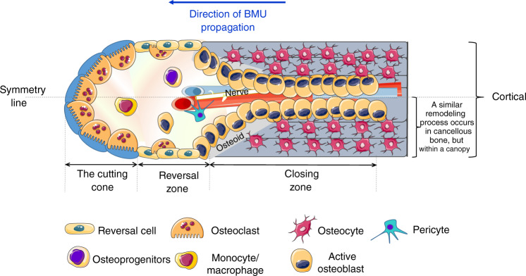
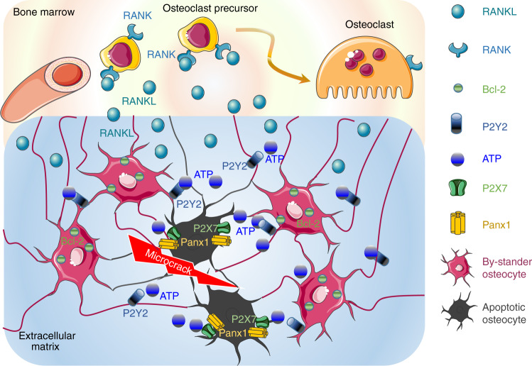
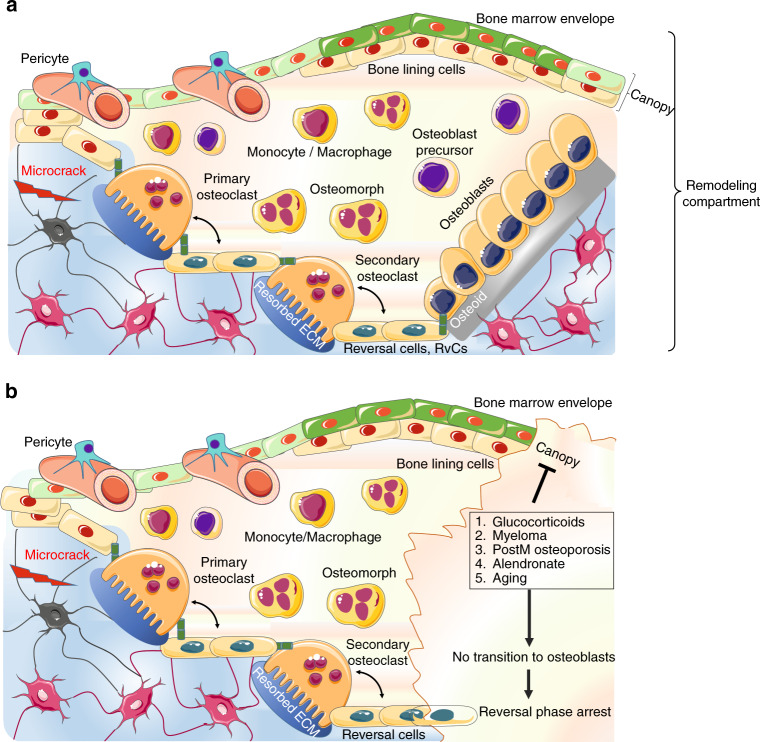
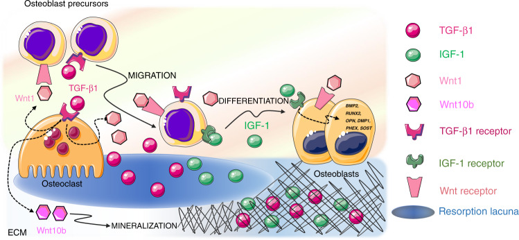
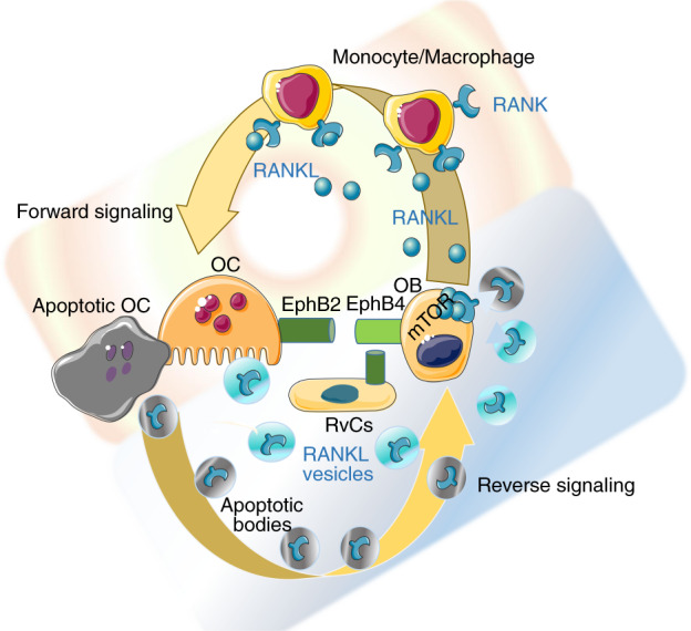
Bone remodeling replaces old and damaged bone with new bone through a sequence of cellular events occurring on the same surface without any change in bone shape. It was initially thought that the basic multicellular unit (BMU) responsible for bone remodeling consists of osteoclasts and osteoblasts functioning through a hierarchical sequence of events organized into distinct stages. However, recent discoveries have indicated that all bone cells participate in BMU formation by interacting both simultaneously and at different differentiation stages with their progenitors, other cells, and bone matrix constituents. Therefore, bone remodeling is currently considered a physiological outcome of continuous cellular operational processes optimized to confer a survival advantage. Bone remodeling defines the primary activities that BMUs need to perform to renew successfully bone structural units. Hence, this review summarizes the current understanding of bone remodeling and future research directions with the aim of providing a clinically relevant biological background with which to identify targets for therapeutic strategies in osteoporosis.


Conflict of interest statement

The authors declare no competing interests.

Figures

Fig. 1
Fig. 1
The cutting cone. The cutting cone originates in close proximity to neurovascular axial bundles and is generated by the propagation of the basic multicellular unit (BMU). The cutting cone consists of a set of osteoclasts, followed by a set of osteoblasts, reversal cells and secondary osteoclasts that cover the so-called reversal zone. At the end of the reversal zone, a set of osteocytes generate the closing zone. A line of symmetry divides in half the representation of a complete BMU in the cortex moving toward the longitudinal axis of the long bone. One-half of cortical BMU is similar to the BMU at the cancellous surface, although in cancellous bone, the BMU is separated from the marrow by a specific cell structure called the canopy
Fig. 2
Fig. 2
The “find me” message from apoptotic osteocytes. Under fatigue failure, osteocytes undergo apoptosis, whereas the osteocytes surrounding the apoptotic osteocytes, the “bystander osteocytes”, show upregulation of the antiapoptotic protein BcL-2, which protects them from death. Apoptotic osteocytes activate the Panx1/P2XR pathway to induce the release of ATP into the extracellular compartment as a specific “find me” signal. ATP binds P2Y2 receptors on bystander osteocytes, which in turn produce and release RANKL at the interface with the bone marrow. RANKL promotes the recruitment of osteoclast progenitors and osteoclastogenesis
Fig. 3
Fig. 3
The bone remodeling compartment (BRC). a The BRC provides the correct microenvironment to link bone formation and resorption through local signaling. The bone marrow envelope (BME) is a layer of cells of mesenchymal origin and a reservoir for osteoprogenitors that covers the layer of bone lining cells (BLCs). Once remodeling is initiated, osteoclasts lift the BME from the BLC, inducing BME cells to form a structure called the canopy. The canopy separates the remodeling site from the remainder of the bone marrow to allow osteoclast and osteoblast precursors to enter the blood compartment. After resorption, osteoclasts either undergo apoptosis or dedifferentiate into osteomorphs. The resorbed surface is then colonized by secondary osteoclasts and reversal cells. Reversal cells are osteoblast progenitors that digest fibrillar collagen remnants, similar to BLCs. Secondary osteoclasts and reversal cells provide the basis for the recruitment and expansion of osteoblastic pools (b) except under certain counteracting conditions, such as glucocorticoid or alendronate treatment, myeloma, or postmenopausal osteoporosis
Fig. 4
Fig. 4
Released extracellular matrix (ECM) factors. Two major factors are released by the ECM upon bone resorption and cooperate to regulate bone remodeling and BMU activity: TGF-β1 and IGF-1. After release from the matrix, active TGF-β1 acts both on osteoblasts and osteoclasts to induce osteoblast precursor migration to the site of resorption and osteoclast production of Wnt1, which promotes osteoblast recruitment and/or differentiation at sites of bone resorption, and Wint10b, which promotes matrix mineralization. IGF-1 supports the recruitment of osteoblast progenitors and promotes osteoblast differentiation and matrix mineralization by inducing the transcription of osteogenesis‐related genes such as DMP1, PHEX, SOST, BMP2, RUNX2, OPN, and OCN
Fig. 5
Fig. 5
Anabolic osteoclasts. Osteoblasts stimulate osteoclastogenesis by producing RANKL as a forward signal. However, osteoclasts can act as anabolic cells by generating positive reverse signaling in osteoblasts. Both resorbing and apoptotic osteoclasts can release extracellular vesicles (EVs) that contain RANK. Once discharged from EVs, RANK binds RANKL clusters on the osteoblast membrane and activates osteogenesis via the mTOR pathway. In addition, EphrinB2 signaling from osteoclasts to EphB4 on osteoblasts/bone lining cells (BLCs)/reversal cells favors osteogenic differentiation

References

    1. Frost, H. M. Bone remodeling dynamics (Charles C Thomas Company, 1963).
    1. Delaisse JM, et al. Re-thinking the bone remodeling cycle mechanism and the origin of bone loss. Bone. 2020;141:115628. doi: 10.1016/j.bone.2020.115628. - DOI - PubMed
    1. Maggiano CM, Maggiano IS, Tiesler VG, Chi-Keb JR, Stout SD. Methods and theory in bone modeling drift: comparing spatial analyses of primary bone distributions in the human humerus. J. Anat. 2016;228:190–202. doi: 10.1111/joa.12383. - DOI - PMC - PubMed
    1. Parfitt AM. Osteonal and hemi-osteonal remodeling: the spatial and temporal framework for signal traffic in adult human bone. J. Cell. Biochem. 1994;55:273–286. doi: 10.1002/jcb.240550303. - DOI - PubMed
    1. Frost HM. A synchronous group of mammalian cells whose in vivo behavior can be studied. Henry Ford. Hosp. Med. Bull. 1965;13:161–172. - PubMed