. 2022 Sep;36(9):2281-2292.
doi: 10.1038/s41375-022-01641-x. Epub 2022 Jul 18.

PARP14 is a novel target in STAT6 mutant follicular lymphoma

Affiliations

PARP14 is a novel target in STAT6 mutant follicular lymphoma

Michael Mentz et al. Leukemia. 2022 Sep.

Abstract

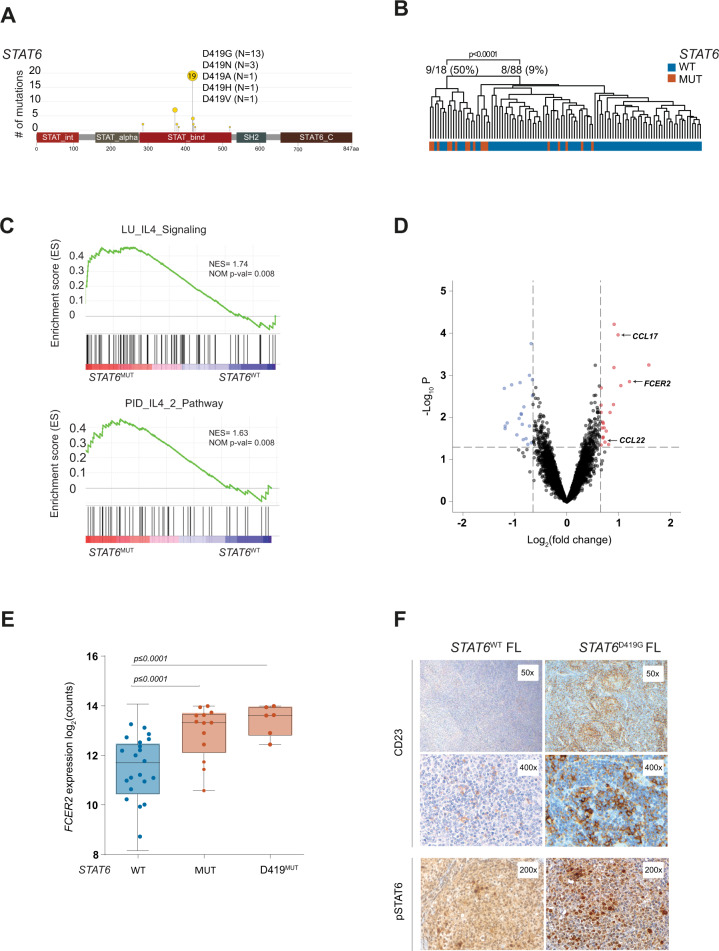
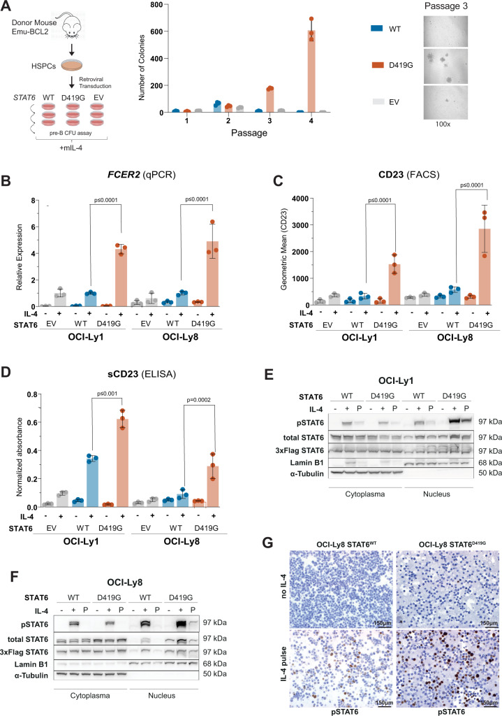
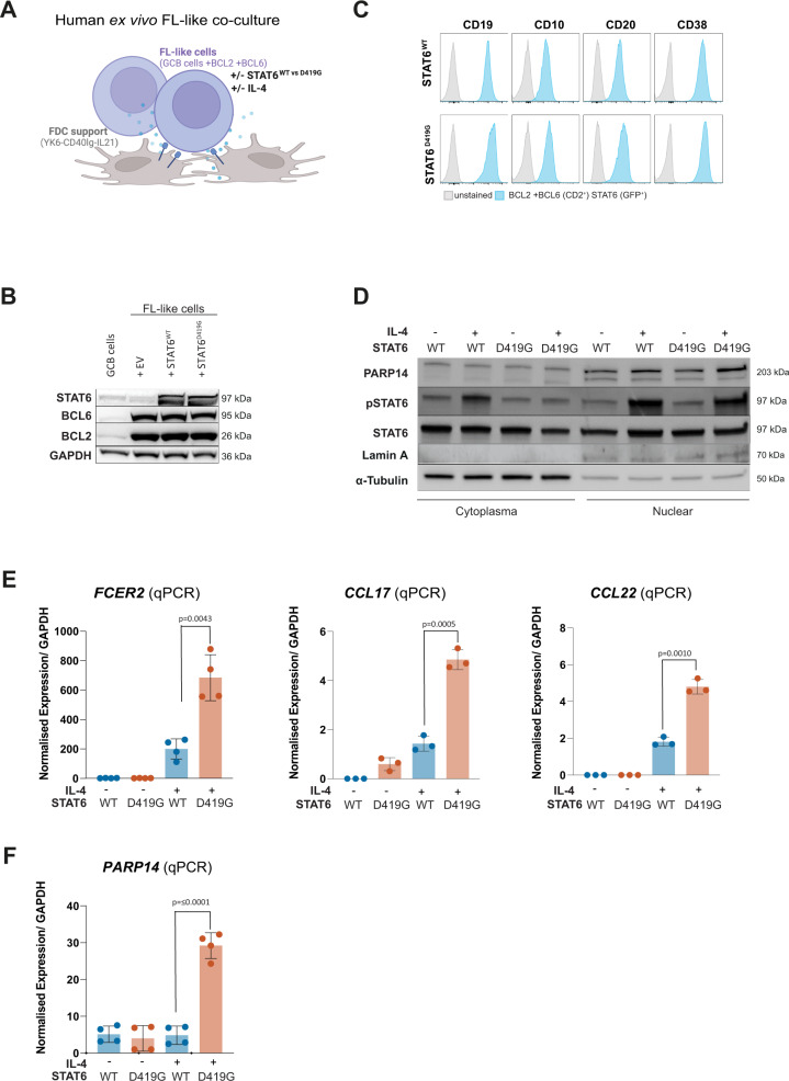
The variable clinical course of follicular lymphoma (FL) is determined by the molecular heterogeneity of tumor cells and complex interactions within the tumor microenvironment (TME). IL-4 producing follicular helper T cells (TFH) are critical components of the FL TME. Binding of IL-4 to IL-4R on FL cells activates JAK/STAT signaling. We identified STAT6 mutations (STAT6MUT) in 13% of FL (N = 33/258), all clustered within the DNA binding domain. Gene expression data and immunohistochemistry showed upregulation of IL-4/STAT6 target genes in STAT6MUT FL, including CCL17, CCL22, and FCER2 (CD23). Functionally, STAT6MUT was gain-of-function by serial replating phenotype in pre-B CFU assays. Expression of STAT6MUT enhanced IL-4 induced FCER2/CD23, CCL17 and CCL22 expression and was associated with nuclear accumulation of pSTAT6. RNA sequencing identified PARP14 -a transcriptional switch and co-activator of STAT6- among the top differentially upregulated genes in IL-4 stimulated STAT6MUT lymphoma cells and in STAT6MUT primary FL cells. Quantitative chromatin immunoprecipitation (qChIP) demonstrated binding of STAT6MUT but not STAT6WT to the PARP14 promotor. Reporter assays showed increased IL-4 induced transactivation activity of STAT6MUT at the PARP14 promotor, suggesting a self-reinforcing regulatory circuit. Knock-down of PARP14 or PARP-inhibition abrogated the STAT6MUT gain-of-function phenotype. Thus, our results identify PARP14 as a novel therapeutic target in STAT6MUT FL.


Conflict of interest statement

This research was supported by the Deutsche Forschungsgemeinschaft (DFG, German Research Foundation), Project-ID WE 4679/2-1 (OW), SCHM2440/8-1 (MSS), and the Max-Eder Program of the Deutsche Krebshilfe e.V. (DKH, German Cancer Aid), Project-ID 110659/70112997 (OW). OW is supported by an Else-Kröner Excellence Fellowship from the Else-Kröner-Fresenius Stiftung (Project-ID 2021_EKES.13). LA is supported by the Else Kröner-Fresenius Clinician Scientist Program: Cancer immunotherapy, and the Munich Clinician Scientist Program (MCSP). LA and JAH are supported by the German Consortium for Translational Cancer Research (DKTK) School of Oncology. MS, Amgen, Speakers Bureau. The authors declare no competing interests.

Figures

Fig. 1
Fig. 1. STAT6 mutations are associated with activated IL-4 signaling in human FL.
A Lolliplot of STAT6 mutations in 258 human FL biopsies. B Unsupervised hierarchical clustering of gene expression data from 106 human FL, annotated for STAT6 genotype. C Same data used for gene set enrichment analysis (GSEA) for established IL-4 signatures comparing STAT6WT and STAT6MUT. D Volcano plot of differentially expressed genes between STAT6WT and STAT6MUT FL (N = 106, log2FC = ±0.65, p value = 0.05). E FCER2 (CD23) gene expression in human FL with wild type STAT6 (STAT6WT) vs any STAT6 mutation (STAT6MUT) vs STAT6 mutations at position D419 (STAT6D419). F Representative immunohistochemistry (IHC) stains for CD23 and pSTAT6 in human FL with STAT6WT (left) and STAT6MUT (STAT6D419G, right).
Fig. 2
Fig. 2. STAT6 DNA binding site mutations are gain-of-function and require IL-4 for increased STAT6 activation.
A Hematopoietic stem and progenitor cells (HSPCs) from Emu-BCL2 mouse cells transduced with either STAT6WT, STAT6D419G or empty vector (EV) and serially replated on cytokine-supplemented methylcellulose (MethoCult, M3630) that supports the growth of mouse pre-B colony-forming units (CFUs) plus additional mouse IL-4 (mIL-4) (N = 3, mean ± SD). B FCER2 mRNA levels (by qPCR) in OCI-Ly1 and OCI-Ly8 cells expressing either STAT6WT, STAT6D419G or EV control after IL-4 stimulation (10 ng/mL, 24 h; 2−dCt values relative to STAT6WT, N = 3, mean ± SD). C CD23 cell surface expression (by FACS) on OCI-Ly1 and OCI-Ly8 expressing either STAT6WT, STAT6D419G or EV control after IL-4 stimulation (10 ng/mL, 24 h; geometric mean, N = 3, mean ± SD). D Soluble CD23 (sCD23) levels (by ELISA) in cell culture supernatants of OCI-Ly1 and OCI-Ly8 cells expressing either STAT6WT, STAT6D419G or EV control after IL-4 stimulation (10 ng/mL, 72 h by ELISA (N = 3, mean ± SD). E Immunoblots of subcellular fractions (cytoplasmic vs nuclear) of OCI-Ly1 and F OCI-Ly8 cells expressing either STAT6WT or STAT6D419G. “−“ indicates no IL-4 stimulation, “+” indicates IL-4 stimulation (10 ng/mL for 20 min), and “P” indicates IL-4 pulse stimulation (IL-4 10 ng/mL for 20 min, then wash & withdrawal of IL-4 and incubation for another 8 h in fresh media without IL-4). G Representative immunohistochemistry (IHC) stain for phosphorylated STAT6 (pSTAT6) in OCI-Ly1 cells expressing STAT6WT or STAT6D419G with or without IL-4 pulse stimulation.
Fig. 3
Fig. 3. PARP14 is strongly upregulated in IL-4-stimulated STAT6MUT lymphoma cells.
A Volcano plot of differentially expressed genes from whole transcriptome sequencing of OCI-Ly1 cells expressing STAT6WTor STAT6MUT (D419G, D419N, N421K) across all time points (i.e., 2, 4, and 8 h, respectively). Dashed lines indicate log2FC ±1.0, adj. p value 0.0001. B PARP14 expression validated by quantitative PCR (qPCR) in OCI-Ly1 cells. C Immunoblot analysis of PARP14 protein expression in OCI-Ly1 and OCI-Ly8 expressing STAT6WT or STAT6D419G after IL-4 stimulation (10 ng/mL, 24 h). D Immunoprecipitation of 3xFlag-tagged STAT6 and immunoblotting for PARP14 and STAT6 (3xFlag) in OCI-LY8 after IL-4 stimulation (10 ng/mL, 24 h). E Proximity ligation assay (PLA) of STAT6 and PARP14. Representative images of OCI-Ly8 cells expressing STAT6WT or STAT6D419G after IL-4 stimulation (left); TexasRed channel used to detect red amplification signal. Box plots display the percentage of TexasRed positive cells per total cell number for each analyzed microscopic view field (N = 5, right). F Single-cell RNA sequencing analysis. Violin plot showing PARP14 expression in tumor cells from 8 FL patients with wild type STAT6 (STAT6WT, N = 6) vs any STAT6 mutation (MUT, N = 2) vs STAT6 mutation at position D419 (D419MUT, N = 1).
Fig. 4
Fig. 4. Validation experiments in a human ex vivo FL-like co-culture system.
A Schematic of a fully human FL-like model system: human tonsil-derived germinal center (GC) B cells expressing BCL2 and BCL6 as well as STAT6WT or STAT6D419G or EV are co-cultured with follicular dendritic cell (FDC) feeder cells that express CD40L and IL-21 (YK6-CD40lg-IL-21). B Western Blot of FL-like cells, whole cell lysates. C Flow cytometry of GC markers on FL-like cells with indicated STAT6 genotypes. D Western Blot of FL-like cells, cellular and nuclear fraction. E qPCR analysis of FCER2, CCL17, and CCL21, as well as F PARP14 in FL-like cells with indicated STAT6 genotypes with and without IL-4 stimulation.
Fig. 5
Fig. 5. PARP14 per se is a target gene of mutant (STAT6MUT) but not wild type STAT6 (STAT6WT).
A Cross-linked chromatin immunoprecipitation (ChIP) of 3xFlag-tagged STAT6WT or STAT6D419G in OCI-LY8 cells after IL-4 stimulation (10 ng/mL, 24 h) followed by quantitative PCR for the PARP14 promoter region (qChIP). B Schematic of the PARP14 promotor region, indicating predicted / putative STAT6 binding sites and the 641 bp region that was cloned into pGL3. C PARP14 promoter luciferase assay (pGL3) with increasing amounts of co-transfected STAT6WT or STAT6D419G in 293 T cells in the presence of IL-4 (24 h after transfection, 10 ng/mL for 6 h). Shown are fold changes (FC) of luciferase activity (relative light unit, RLU) normalized to 1 ng STAT6WT (N = 6, mean ± SD). D CD23 cell surface expression (by FACS) on OCI-Ly1 and E OCI-Ly8, each expressing either STAT6WT or STAT6D419G with shRNA-mediated knock-down of PARP14 (sh4) or a non-targeting (scrambled) control (scr), respectively. Immunoblot of respective cells as indicated below. F CD23 cell surface expression by FACS on OCI-Ly1 and OCI-L8 expressing STAT6WT or STAT6D419G with or without IL-4 stimulation (10 mg/mL, 24 h) and treatment with the PARP inhibitor PJ34 (50 µM, 15 min prior to IL-4 stimulation) or vehicle, respectively. (N = 3, mean ± SD). Cell viability of cells as indicated below (N = 3, mean ± SD).
Fig. 6
Fig. 6. Model of a PARP14-mediated self-reinforcing regulatory circuit that amplifies IL-4 induced transcriptional activity in STAT6MUT FL.
STAT6 mutations amplify IL-4 induced STAT6-dependent gene activation via an intracellular self-reinforcing regulatory microcircuit that involves aberrantly increased PARP14 levels in IL-4 stimulated STAT6MUT FL cells. Increased STAT6-dependent gene expression involves cytokines (e.g., CCL17 and CCL22) which contribute to the re-education of the tumor microenvironment, including increased recruitment of IL-4 producing T follicular helper (TFH) cells. Details see text. Yellow star indicates STAT6 mutation. Figure created with BioRender.com.

References

    1. Hiddemann W, Cheson BD. How we manage follicular lymphoma. Leukemia. 2014;28:1388–95. doi: 10.1038/leu.2014.91. - DOI - PubMed
    1. Casulo C, Byrtek M, Dawson KL, Zhou X, Farber CM, Flowers CR, et al. Early Relapse of Follicular Lymphoma After Rituximab Plus Cyclophosphamide, Doxorubicin, Vincristine, and Prednisone Defines Patients at High Risk for Death: An Analysis From the National LymphoCare Study. J Clin Oncol : Off J Am Soc Clin Oncol. 2015;33:2516–22. doi: 10.1200/JCO.2014.59.7534. - DOI - PMC - PubMed
    1. Jurinovic V, Kridel R, Staiger AM, Szczepanowski M, Horn H, Dreyling MH, et al. Clinicogenetic risk models predict early progression of follicular lymphoma after first-line immunochemotherapy. Blood. 2016;128:1112–20. doi: 10.1182/blood-2016-05-717355. - DOI - PMC - PubMed
    1. Carbone A, Roulland S, Gloghini A, Younes A, von Keudell G, López-Guillermo A, et al. Follicular lymphoma. Nat Rev Dis Prim. 2019;5:83. doi: 10.1038/s41572-019-0132-x. - DOI - PubMed
    1. Kridel R, Sehn LH, Gascoyne RD. Pathogenesis of follicular lymphoma. J Clin Investig. 2012;122:3424–31.. doi: 10.1172/JCI63186. - DOI - PMC - PubMed

Publication types