

Characterization of circumferential antral pulmonary vein isolation areas resulting from pulsed-field catheter ablation
- PMID: 35852306
- PMCID: PMC10103571
- DOI: 10.1093/europace/euac111
Characterization of circumferential antral pulmonary vein isolation areas resulting from pulsed-field catheter ablation
Abstract
Aims: The cornerstone of pulmonary vein (PV) isolation (PVI) is a wide-area circumferential ablation (WACA) resulting in an antral PVI area. Pulsed-field ablation (PFA) is a new nonthermal 'single-shot' PVI technique resulting in well-characterized posterior isolation areas. However, information on circumferential PVI area is lacking. Thus, we sought to characterize the circumferential antral PVI areas after PFA-PVI.
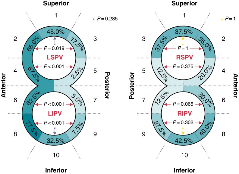
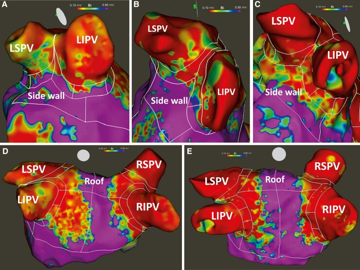
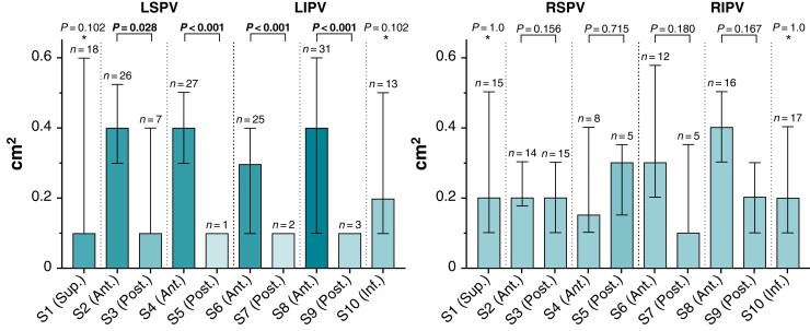
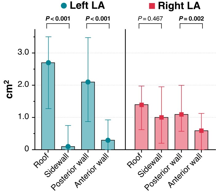
Methods and results: Atrial fibrillation (AF) patients underwent fluoroscopy-guided PVI with a pentaspline PFA catheter. Ultra-high-density voltage maps using a 20-polar circular mapping catheter were created before and immediately after PVI to identify and quantify (i) insufficient isolation areas per antral PV segment (10-segment model) and (ii) enlarged left atrial (LA) isolation areas (beyond the antral PV segments) per LA region (8-region model). The PFA-PVI with pre- (5469 ± 1822 points) and post-mapping (6809 ± 2769 points) was performed in 40 consecutive patients [age 62 ± 6 years, 25/40 (62.5%) paroxysmal AF]. Insufficient isolation areas were located most frequently in the anterior antral PV segments of the left PVs (62.5-77.5% of patients) with the largest extent (median ≥0.4 cm2) located in the same segments (segments 2/5/8). Enlarged LA isolation areas were located most frequently and most extensively on the posterior wall and roof region (89.5-100% of patients; median 1.1-2.7 cm2 per region).
Conclusion: Fluoroscopy-guided PFA-PVI frequently results in insufficient isolation areas in the left anterior antral PV segments and enlarged LA isolation areas on the posterior wall/roof, which both may be extensive. To optimize the procedure, full integration of PFA catheter visualization into three-dimensional-mapping systems is needed.
Keywords: Antral; Catheter ablation; Circumferential; Isolation area; Pulmonary vein isolation; Pulsed-field ablation.
© The Author(s) 2022. Published by Oxford University Press on behalf of the European Society of Cardiology. All rights reserved. For permissions, please email: journals.permissions@oup.com.
Conflict of interest statement
Conflict of interest: M.B. reports educational grant support from Boston Scientific (Fellowship ‘Herzrhythmus’). F.J.N. reports lecture fees paid to his institution from Amgen, Bayer Healthcare, Boehringer Ingelheim, Boston Scientific, Daiichi Sankyo, Edwards Lifesciences, Ferrer, Pfizer, Novartis; consultancy fees paid to his institution from Boehringer Ingelheim, Novartis, and grant support from Bayer Healthcare, Boston Scientific, Biotronik, Edwards Lifesciences, GlaxoSmithKline, Medtronic, Pfizer, Abbot Vascular. All the remaining authors have declared no conflicts of interest.
Figures
References
-
- Hindricks G, Potpara T, Dagres N, Arbelo E, Bax JJ, Blomström-Lundqvist Cet al. 2020 ESC Guidelines for the diagnosis and management of atrial fibrillation developed in collaboration with the European Association for Cardio-Thoracic Surgery (EACTS). Eur Heart J 2021;42:373–498. - PubMed
-
- Arentz T, Weber R, Bürkle G, Herrera C, Blum T, Stockinger Jet al. Small or large isolation areas around the pulmonary veins for the treatment of atrial fibrillation? Results from a prospective randomized study. Circulation 2007;115:3057–63. - PubMed
-
- Kiuchi K, Kircher S, Watanabe N, Gaspar T, Rolf S, Arya Aet al. Quantitative analysis of isolation area and rhythm outcome in patients with paroxysmal atrial fibrillation after circumferential pulmonary vein antrum isolation using the pace-and-ablate technique. Circ Arrhythmia Electrophysiol 2012;5:667–75. - PubMed
-
- Chierchia GB, De AC, Sorgente A, Paparella G, Sarkozy A, Müller-Burri SAet al. Anatomical extent of pulmonary vein isolation after cryoballoon ablation for atrial fibrillation: comparison between the 23 and 28 mm balloons. J Cardiovasc Med 2011;12:162–6. - PubMed
-
- Kenigsberg DN, Martin N, Lim HW, Kowalski M, Ellenbogen KA. Quantification of the cryoablation zone demarcated by pre- and postprocedural electroanatomic mapping in patients with atrial fibrillation using the 28-mm second-generation cryoballoon. Heart Rhythm 2015;12:283–90. - PubMed
MeSH terms
LinkOut - more resources
Full Text Sources
Medical
Research Materials
