. 2022 Aug 31;10(4):e0025022.
doi: 10.1128/spectrum.00250-22. Epub 2022 Jul 19.

Rhamnolipid-Coated Iron Oxide Nanoparticles as a Novel Multitarget Candidate against Major Foodborne E. coli Serotypes and Methicillin-Resistant S. aureus

Affiliations

Rhamnolipid-Coated Iron Oxide Nanoparticles as a Novel Multitarget Candidate against Major Foodborne E. coli Serotypes and Methicillin-Resistant S. aureus

Mohamed Sharaf et al. Microbiol Spectr. .

Abstract

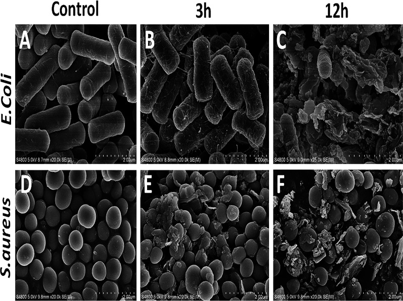
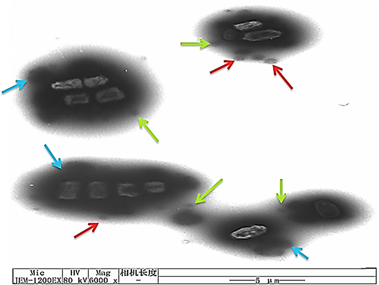
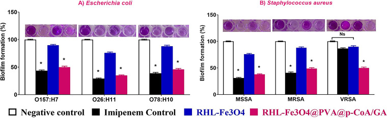
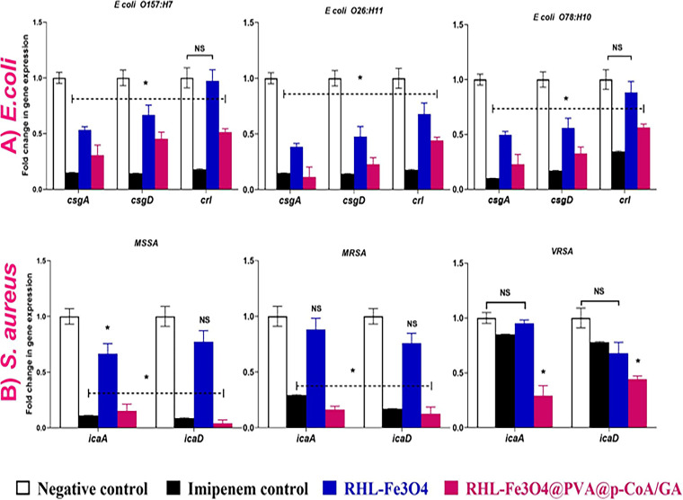
Surface-growing antibiotic-resistant pathogenic bacteria such as Escherichia coli and Staphylococcus aureus are emerging as a global health challenge due to dilemmas in clinical treatment. Furthermore, their pathogenesis, including increasingly serious antimicrobial resistance and biofilm formation, makes them challenging to treat by conventional therapy. Therefore, the development of novel antivirulence strategies will undoubtedly provide a path forward in combatting these resistant bacterial infections. In this regard, we developed novel biosurfactant-coated nanoparticles to combine the antiadhesive/antibiofilm properties of rhamnolipid (RHL)-coated Fe3O4 nanoparticles (NPs) with each of the p-coumaric acid (p-CoA) and gallic acid (GA) antimicrobial drugs by using the most available polymer common coatings (PVA) to expand the range of effective antibacterial drugs, as well as a mechanism for their synergistic effect via a simple method of preparation. Mechanistically, the average size of bare Fe3O4 NPs was ~15 nm, while RHL-coated Fe3O4@PVA@p-CoA/GA was about ~254 nm, with a drop in zeta potential from -18.7 mV to -34.3 mV, which helped increase stability. Our data show that RHL-Fe3O4@PVA@p-CoA/GA biosurfactant NPs can remarkably interfere with bacterial growth and significantly inhibited biofilm formation to more than 50% via downregulating IcaABCD and CsgBAC operons, which are responsible for slime layer formation and curli fimbriae production in S. aureus and E. coli, respectively. The novelty regarding the activity of RHL-Fe3O4@PVA@p-CoA/GA biosurfactant NPs reveals their potential effect as an alternative multitarget antivirulence candidate to minimize infection severity by inhibiting biofilm development. Therefore, they could be used in antibacterial coatings and wound dressings in the future. IMPORTANCE Antimicrobial resistance poses a great threat and challenge to humanity. Therefore, the search for alternative ways to target and eliminate microbes from plant, animal, and marine microorganisms is one of the world's concerns today. Furthermore, the extraordinary capacity of S. aureus and E. coli to resist standard antibacterial drugs is the dilemma of all currently used remedies. Methicillin-resistant S. aureus (MRSA) and vancomycin-resistant S. aureus (VRSA) have become widespread, leading to no remedies being able to treat these threatening pathogens. The most widely recognized serotypes that cause severe foodborne illness are E. coli O157:H7, O26:H11, and O78:H10, and they display increasing antimicrobial resistance rates. Therefore, there is an urgent need for an effective therapy that has dual action to inhibit biofilm formation and decrease bacterial growth. In this study, the synthesized RHL-Fe3O4@PVA@p-CoA/GA biosurfactant NPs have interesting properties, making them excellent candidates for targeted drug delivery by inhibiting bacterial growth and downregulating biofilm-associated IcaABCD and CsgBAC gene loci.

Keywords: antiadhesive property; antimicrobial activity; biofilm formation; drug delivery; iron oxide nanoparticles; rhamnolipids.


Conflict of interest statement

The authors declare no conflict of interest.

Figures

FIG 1
FIG 1
(A) Schematic illustration of prepared RHL-Fe3O4@PVA@p-CoA/GA biosurfactant magnetic NPs according to the emulsion-coacervation method at 20 to 22°C. (B) Exchange of chemosorbed rhamnolipid (RHL) ligands on the Fe3O4@PVA@p-CoA/GA NP surfaces via hydrogen binding donor. (C to E) FTIR spectra (C), XRD pattern (D), and VSM analysis (E) of bare Fe3O4 and Fe3O4@PVA@p-CoA/GA NPs and RHL-Fe3O4@PVA@p-CoA/GA biosurfactant NPs.
FIG 2
FIG 2
Average particle size and size distribution of prepared samples. (A and D) Bare Fe3O4 (scale bar, 100 nm); (B and E) Fe3O4@PVA@p-CoA/GA NPs (scale bar, 100 nm); (C and F) RHL-Fe3O4@PVA@p-CoA/GA NPs (scale bar, 50 nm) measured by TEM. Data of size distribution are presented as means ± SD (n =3).
FIG 3
FIG 3
SEM analysis of prepared NPs. (A to C) Bare Fe3O4 (A), Fe3O4@PVA@p-CoA/GA (B), and RHL-Fe3O4@PVA@p-CoA/GA (C) NPs (scale bar, 200 nm).
FIG 4
FIG 4
In vitro drug release evaluation shown as the percentage of cumulative release and TEM. (A) Release profile of both p-CoA and GA for prepared RHL-Fe3O4@PVA@p-CoA/GA NPs at pH 1.2, 6.8, and 7.4 at 37°C. (B) TEM images of the change in the shape and size of RHL-Fe3O4@PVA@p-CoA/GA NPs with release of the drugs at pH values of 1.2, 6.8, and 7.4 after 3, 5, and 48 h, respectively, at 37°C (scale bar, 2 μm).
FIG 5
FIG 5
RHL-Fe3O4 and RHL-Fe3O4@PVA@p-CoA/GA NPs inhibit E. coli and S. aureus bacterial growth. Inhibitory effects of RHL-Fe3O4 and RHL-Fe3O4@PVA@p-CoA/GA NPs compared with imipenem and vancomycin as standard antibiotic control were determined in vitro by agar well diffusion assay. (A) E. coli O157:H7, O26:H11, and O78:H10; (B) MSSA, MRSA, and VRSA. Each column shows the mean ± SD of three independent experiments, and representative images are shown. Asterisk represents statistically significant differences (P < 0.05), and “ns” represents nonsignificant differences (P > 0.05) compared to the control sample.
FIG 6
FIG 6
SEM images of E. coli and S. aureus after 3 and 12 h exposure to the different treatments. (A and D) Untreated (control); (B and E) treatment with MICs of RHL-Fe3O4@PVA@p-CoA/GA biosurfactant NPs after 3 h; (C and F) treatment with MICs of RHL-Fe3O4@PVA@p-CoA/GA biosurfactant NPs after12 h (scale bar, 2 μm).
FIG 7
FIG 7
Mechanism of action between RHL-Fe3O4@PVA@p-CoA/GA biosurfactant NPs and E. coli bacteria cell membrane, measured by TEM (scale bar, 5 μm).
FIG 8
FIG 8
RHL-Fe3O4 and RHL-Fe3O4@PVA@p-CoA/GA NPs reduce the initial adhesion and biofilm formation of E. coli O157:H7, O26:H11, and O78:H10 (A) and S. aureus MSSA, MRSA, and VRSA (B). The biofilms of treated bacteria were detected by crystal violet staining and quantified by measuring the OD600. Each column shows the mean ± SD of three independent experiments, and representative images are shown; increasing violet color indicates higher biofilm formation. Asterisk represents statistically significant differences (P < 0.05), and “ns” represents nonsignificant differences (P > 0.05) compared to the control sample.
FIG 9
FIG 9
Transcriptional profile of biofilm-associated genes upon treatment with RHL-Fe3O4@PVA@p–CoA/GA NPs. (A) E. coli O157:H7, O26:H11, and O78:H10; (B) MSSA, MRSA, and VRSA. Relative gene expression levels of csgA, csgD, crl, icaA, and icaD were calculated using the ΔΔCT method and expressed as fold change. 16S rRNA was used as the endogenous control. Each column shows the mean ± SD of three independent experiments Asterisk represents statistically significant differences (P < 0.05), and “ns” represents nonsignificant differences (P > 0.05) compared to the control sample.
FIG 10
FIG 10
Proposed mechanistic illustration of multitarget activity of RHL-Fe3O4@PVA@p-CoA/GA biosurfactant NPs of suppression/inhibition on the plankton bacteria-biofilm interface and the activation of the bacterial cell death signaling cascade. ONPG, o-nitrophenyl-β-d-galactopyranoside.

References

    1. Tong SY, Davis JS, Eichenberger E, Holland TL, Fowler VG, Jr.. 2015. Staphylococcus aureus infections: epidemiology, pathophysiology, clinical manifestations, and management. Clin Microbiol Rev 28:603–661. doi:10.1128/CMR.00134-14. - DOI - PMC - PubMed
    1. Mendes RE, Mendoza M, Banga Singh KK, Castanheira M, Bell JM, Turnidge JD, Lin SSF, Jones RN. 2013. Regional resistance surveillance program results for 12 Asia-Pacific nations (2011). Antimicrob Agents Chemother 57:5721–5726. doi:10.1128/AAC.01121-13. - DOI - PMC - PubMed
    1. Wunderink RG, Rello J, Cammarata SK, Croos-Dabrera RV, Kollef MH. 2003. Linezolid vs vancomycin: analysis of two double-blind studies of patients with methicillin-resistant Staphylococcus aureus nosocomial pneumonia. Chest 124:1789–1797. doi:10.1016/S0012-3692(15)33412-7. - DOI - PubMed
    1. Gould I. 2013. Treatment of bacteraemia: meticillin-resistant Staphylococcus aureus (MRSA) to vancomycin-resistant S. aureus (VRSA). Int J Antimicrobial Agents 42:S17–S21. doi:10.1016/j.ijantimicag.2013.04.006. - DOI - PubMed
    1. Boll EJ, Struve C, Boisen N, Olesen B, Stahlhut SG, Krogfelt KA. 2013. Role of enteroaggregative Escherichia coli virulence factors in uropathogenesis. Infect Immun 81:1164–1171. doi:10.1128/IAI.01376-12. - DOI - PMC - PubMed

Publication types

LinkOut - more resources