. 2022 Aug 31;10(4):e0101822.
doi: 10.1128/spectrum.01018-22. Epub 2022 Jul 19.

Ability of Two Strains of Lactic Acid Bacteria To Inhibit Listeria monocytogenes by Spot Inoculation and in an Environmental Microbiome Context

Affiliations

Ability of Two Strains of Lactic Acid Bacteria To Inhibit Listeria monocytogenes by Spot Inoculation and in an Environmental Microbiome Context

Priscilla Sinclair et al. Microbiol Spectr. .

Abstract

We evaluated the ability of two strains of lactic acid bacteria (LAB) to inhibit L. monocytogenes using spot inoculation and environmental microbiome attached-biomass assays. LAB strains (PS01155 and PS01156) were tested for antilisterial activity toward 22 phylogenetically distinct L. monocytogenes strains isolated from three fruit packing environments (F1, F2, and F3). LAB strains were tested by spot inoculation onto L. monocytogenes lawns (108 and 107 CFU/mL) and incubated at 15, 20, 25, or 30°C for 3 days. The same LAB strains were also cocultured at 15°C for 3, 5, and 15 days in polypropylene conical tubes with L. monocytogenes and environmental microbiome suspensions collected from F1, F2, and F3. In the spot inoculation assay, PS01156 was significantly more inhibitory toward less concentrated L. monocytogenes lawns than more concentrated lawns at all the tested temperatures, while PS01155 was significantly more inhibitory toward less concentrated lawns only at 15 and 25°C. Furthermore, inhibition of L. monocytogenes by PS01156 was significantly greater at 15°C than higher temperatures, whereas the temperature did not have an effect on the inhibitory activity of PS01155. In the assay using attached environmental microbiome biomass, L. monocytogenes concentration was significantly reduced by PS01156, but not PS01155, when cocultured with microbiomes from F1 and F3 and incubated for 3 days at 15°C. Attached biomass microbiota composition was significantly affected by incubation time but not by LAB strain. This study demonstrates that LAB strains that may exhibit inhibitory properties toward L. monocytogenes in a spot inoculation assay may not maintain antilisterial activity within a complex microbiome. IMPORTANCE Listeria monocytogenes has previously been associated with outbreaks of foodborne illness linked to consumption of fresh produce. In addition to conventional cleaning and sanitizing, lactic acid bacteria (LAB) have been studied for biocontrol of L. monocytogenes in food processing environments that are challenging to clean and sanitize. We evaluated whether two specific LAB strains, PS01155 and PS01156, can inhibit the growth of L. monocytogenes strains in a spot inoculation and in an attached-biomass assay, in which they were cocultured with environmental microbiomes collected from tree fruit packing facilities. LAB strains PS01155 and PS01156 inhibited L. monocytogenes in a spot inoculation assay, but the antilisterial activity was lower or not detected when they were grown with environmental microbiota. These results highlight the importance of conducting biocontrol challenge tests in the context of the complex environmental microbiomes present in food processing facilities to assess their potential for application in the food industry.

Keywords: Listeria monocytogenes; attached biomass; biocontrol; biofilms; environmental microbiome; inhibition; lactic acid bacteria.


Conflict of interest statement

The authors declare no conflict of interest.

Figures
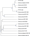
FIG 1
FIG 1
Phylogenetic tree based on and whole-genome sequence for lactic acid bacteria PS01155 and PS01156 as produced by Type (Strain) Genome Server. The branch lengths are scaled in terms of the Genome BLAST Distance Phylogeny (GBDP) distance formula d5. The numbers above branches are GBDP pseudo-bootstrap support values of >60% from 100 replications, with an average branch support of 43.1%. The tree was rooted at the midpoint.
FIG 2
FIG 2
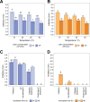
Inhibition of L. monocytogenes strains by (A) PS01155 and (B) PS01156 at 20, 25, and 30°C (n = 22) and at 15°C (n = 21) (n represents the number of L. monocytogenes strains tested at each temperature) using the spot inoculation assay on BHI agar plates. Light bars represent the average zone of inhibition observed on 107CFU/mL L. monocytogenes lawns, and dark bars represent the average zone of inhibition observed on the 108CFU/mL L. monocytogenes lawns at 15, 20, 25, and 30°C, with standard error bars. Average zone of inhibition by supernatants of PS01155 (C) and PS01156 (D) after filtration, pH neutralization, catalase, and proteinase K treatment to determine the nature of the inhibition. Dark bars represent the supernatant obtained from 24-h cultures grown in MRS broth, and light bars represent the supernatant obtained from 48-h cultures grown in MRS broth. For each panel, letters represent significant differences between treatments (P < 0.05) as determined by one-way ANOVA followed by Tukey’s HSD post hoc test.
FIG 3
FIG 3
Aerobic plate counts in the attached biomass grown for 3 (A), 5 (D), and 15 (G) days and L. monocytogenes concentration in the attached biomass grown for 3 (B), 5 (E), and 15 (H) days from environmental microbiomes collected from facilities F1, F2, and F3. NC, negative control; PC, positive control. Bars are color coded by facility, and the error bars represent standard errors. For each panel, letters represent significant differences between treatments (P < 0.05). Microbiota composition of the attached biomass grown for 3 (C), 5 (F), and 15 (I) days. Bars represent the relative abundance of the ASVs with a relative abundance above 2% and are color coded by the assigned taxonomic genus.
FIG 4
FIG 4
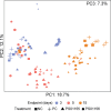
Principal-component analysis plot for the microbiota composition of the attached biomass grown for 3, 5, and 15 days. Each point represents the bacterial composition of one sample. The samples are color coded by growth period, where purple represents the attached-biomass composition for 3-day assay, pink represents a 5-day assay, and orange represents a 15-day assay with repeated culture addition. Squares indicate negative-control (NC) samples, plus symbols indicate positive-control (PC) samples, triangles indicate samples with added PS01155, and circles indicate samples with added PS01156. The size of the symbols represents the third principal component.
FIG 5
FIG 5
Differentially abundant taxa identified in 3-, 5-, and 15-day attached biomass. All samples (i.e., those treated with PS01155 and PS01156 and the positive and negative controls) were merged by day of experiment. The x axis represents the log fold change in relative abundance for ASVs that were differentially abundant (P < 0.05) and had an effect size above 1, calculated using ALDEx2. The color of the bars represents the experimental endpoint with increased relative abundance of each ASV. Each bar has a label corresponding to the assigned taxonomic genus for each ASV.

References

    1. CDC. 2018. Burden of foodborne illness: findings. https://www.cdc.gov/foodborneburden/2011-foodborne-estimates.html. Retrieved 4 June 2020.
    1. Azizoglu RO, Osborne J, Wilson S, Kathariou S. 2009. Role of growth temperature in freeze-thaw tolerance of Listeria spp. Appl Environ Microbiol 75:5315–5320. doi: 10.1128/AEM.00458-09. - DOI - PMC - PubMed
    1. Mazaheri T, Cervantes-Huamán BRH, Bermúdez-Capdevila M, Ripolles-Avila C, Rodríguez-Jerez JJ. 2021. Listeria monocytogenes biofilms in the food industry: is the current hygiene program sufficient to combat the persistence of the pathogen? Microorganisms 9:181. doi: 10.3390/microorganisms9010181. - DOI - PMC - PubMed
    1. Piercey MJ, Hingston PA, Truelstrup Hansen L. 2016. Genes involved in Listeria monocytogenes biofilm formation at a simulated food processing plant temperature of 15°C. Int J Food Microbiol 223:63–74. doi: 10.1016/j.ijfoodmicro.2016.02.009. - DOI - PubMed
    1. dos Reis-Teixeira FB, Alves VF, de Martinis ECP. 2017. Growth, viability and architecture of biofilms of Listeria monocytogenes formed on abiotic surfaces. Braz J Microbiol 48:587–591. doi: 10.1016/j.bjm.2017.01.004. - DOI - PMC - PubMed

Publication types

LinkOut - more resources