. 2022 Aug 22;7(16):e156115.
doi: 10.1172/jci.insight.156115.

The UIP/IPF fibroblastic focus is a collagen biosynthesis factory embedded in a distinct extracellular matrix

Affiliations

The UIP/IPF fibroblastic focus is a collagen biosynthesis factory embedded in a distinct extracellular matrix

Jeremy A Herrera et al. JCI Insight. .

Abstract

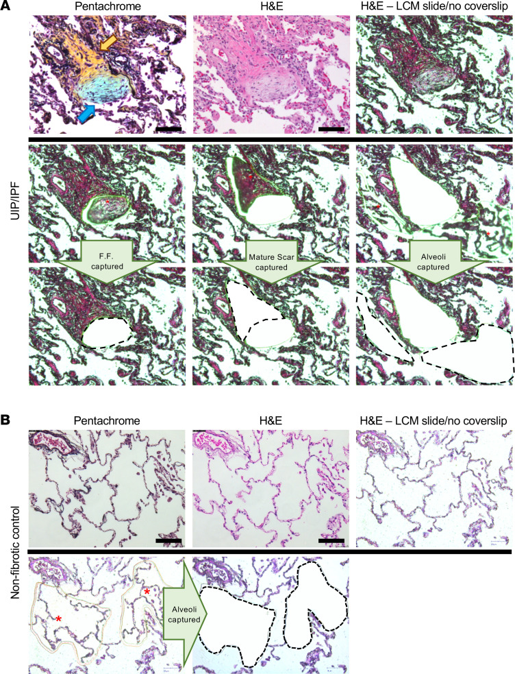
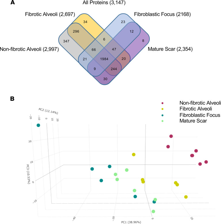
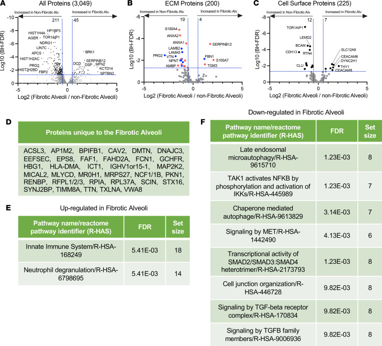
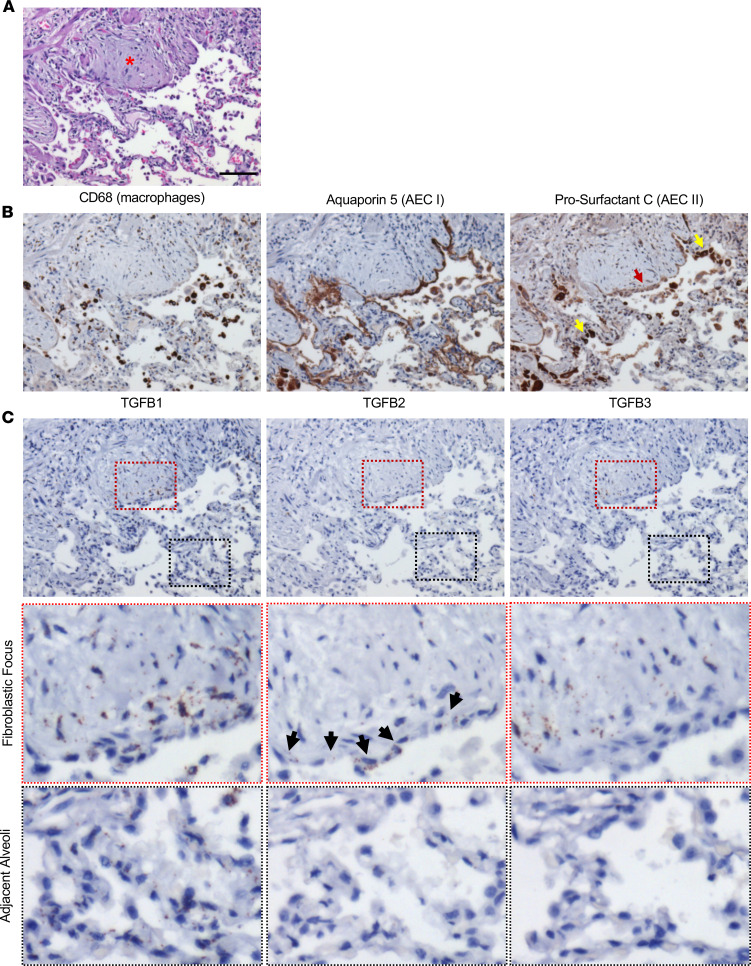
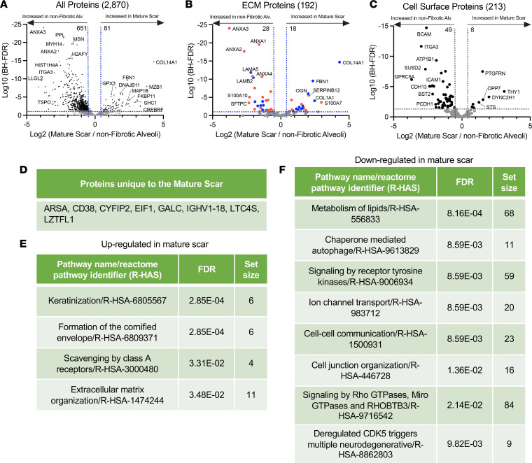
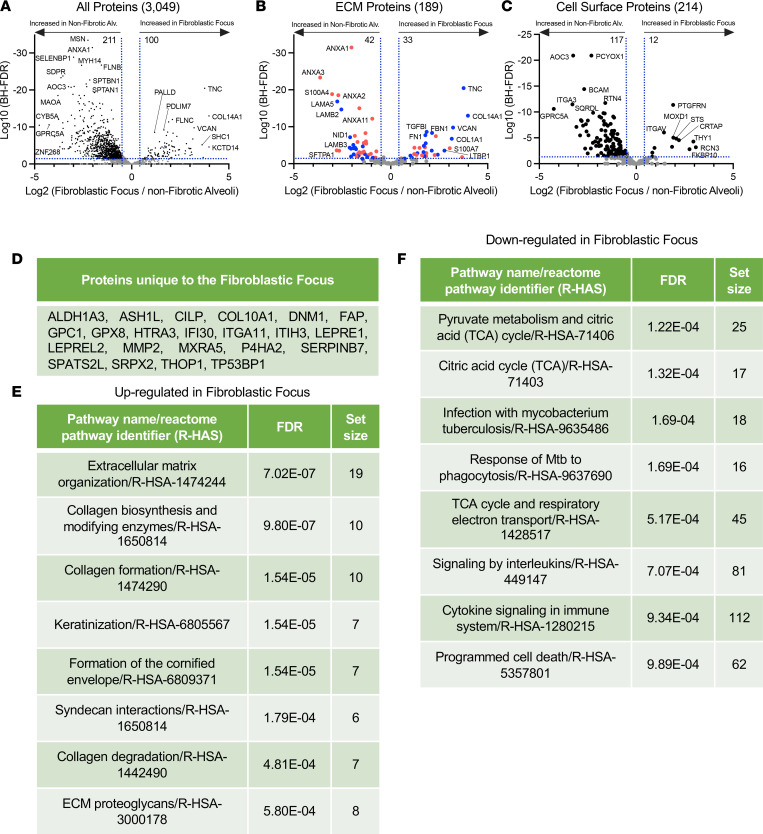
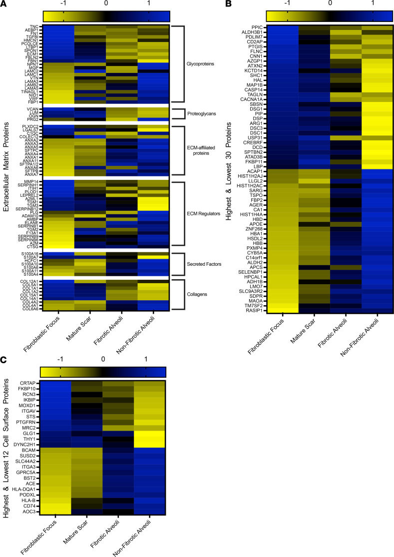
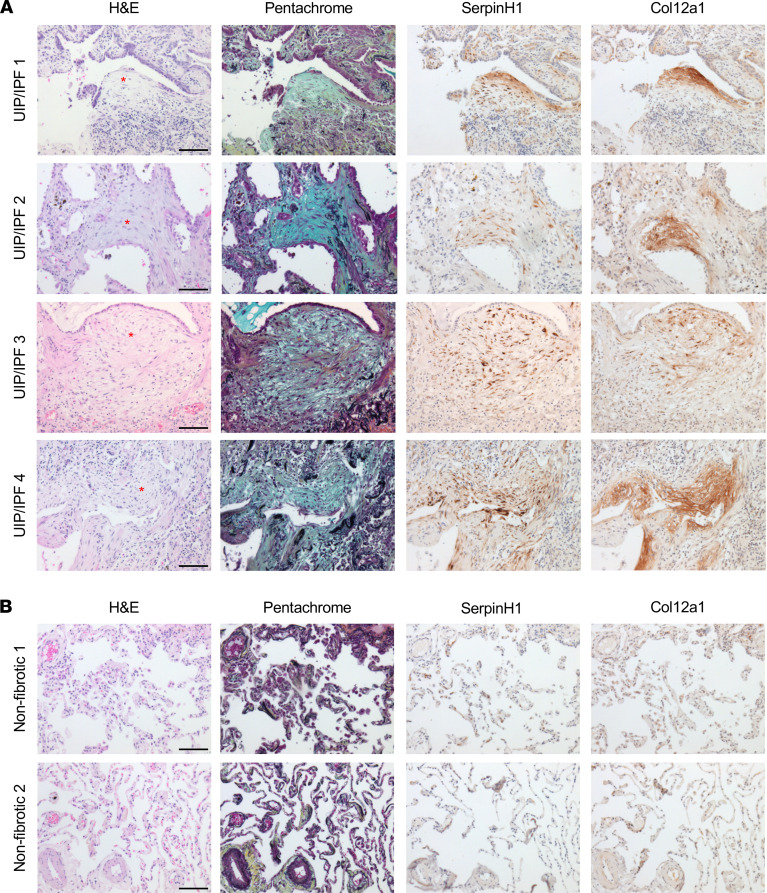
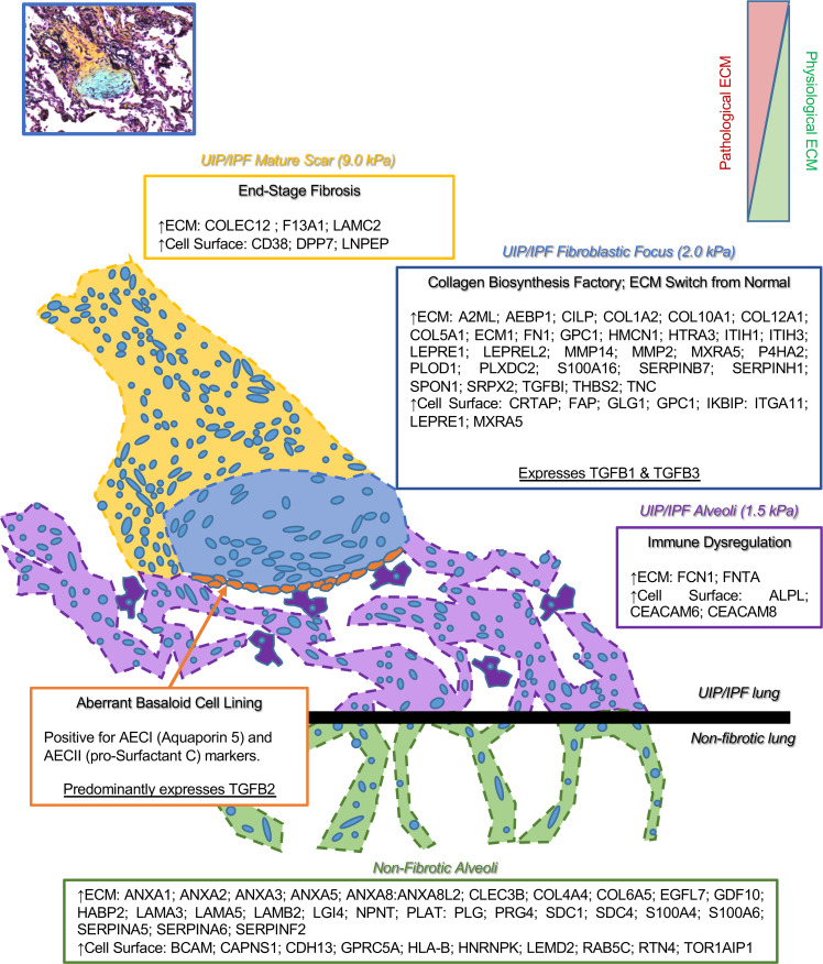
Usual interstitial pneumonia (UIP) is a histological pattern characteristic of idiopathic pulmonary fibrosis (IPF). The UIP pattern is patchy with histologically normal lung adjacent to dense fibrotic tissue. At this interface, fibroblastic foci (FF) are present and are sites where myofibroblasts and extracellular matrix (ECM) accumulate. Utilizing laser capture microdissection-coupled mass spectrometry, we interrogated the FF, adjacent mature scar, and adjacent alveoli in 6 fibrotic (UIP/IPF) specimens plus 6 nonfibrotic alveolar specimens as controls. The data were subjected to qualitative and quantitative analysis and histologically validated. We found that the fibrotic alveoli protein signature is defined by immune deregulation as the strongest category. The fibrotic mature scar classified as end-stage fibrosis whereas the FF contained an overabundance of a distinctive ECM compared with the nonfibrotic control. Furthermore, FF were positive for both TGFB1 and TGFB3, whereas the aberrant basaloid cell lining of FF was predominantly positive for TGFB2. In conclusion, spatial proteomics demonstrated distinct protein compositions in the histologically defined regions of UIP/IPF tissue. These data revealed that FF are the main site of collagen biosynthesis and that the adjacent alveoli are abnormal. This essential information will inform future mechanistic studies on fibrosis progression.

Keywords: Extracellular matrix; Fibrosis; Proteomics; Pulmonology.


Conflict of interest statement

Conflict of interest: The authors have declared that no conflict of interest exists.

Figures

Figure 1
Figure 1. LCM of the FF, mature scar, and adjacent alveoli in a UIP/IPF specimen.
(A) FFPE specimens were serially sectioned at 5 μm and stained with pentachrome (top left) or H&E (the other 8 panels). Notice that pentachrome stains the FF (hallmark lesion in UIP/IPF) in the color blue (blue arrow), while the mature scar tissue appears yellow in color (yellow arrow). We individually captured the FF (left middle and lower panels), the mature scar tissue (mid-middle and lower panels), and the adjacent alveoli (right middle and lower panels) for MS preparation and analysis. (B) Similarly, a nonfibrotic control specimen showing microdissection of alveoli. Scale bar: 100 μm.
Figure 2
Figure 2. Spatial proteomic analysis of UIP/IPF.
UIP/IPF specimens were subjected to LCM-MS to collect mature scar, FF, and fibrotic alveoli (n = 6 UIP/IPF specimens). In addition, LCM-MS was performed to collect alveoli from nonfibrotic controls (n = 6 nonfibrotic specimens). (A) Venn diagram showing all proteins found in each region. (B) A 3-dimensional PCA showing that the FF (dark green dots) is most distant from nonfibrotic alveoli (pink dots), with the mature scar (light green dots) returning toward fibrotic alveoli (yellow dots).
Figure 3
Figure 3. Immune dysregulation defines fibrotic alveoli.
(AC) Volcano plots comparing fibrotic alveoli and nonfibrotic alveoli control showing the negative natural log of the FDR values plotted against the base 2 log (fold change) for each protein. The data in A are for all proteins, whereas data in B were matched against the Human Matrisome Project (34) and C were matched against the Cell Surface Protein Atlas (39). (D) Proteins unique to fibrotic alveoli. Reactome pathways showing the most (E) upregulated or (F) downregulated for fibrotic alveoli compared with nonfibrotic alveoli control.
Figure 4
Figure 4. Distribution of type I and II alveolar epithelial cells and TGFB1–3 in the FF and adjacent alveoli.
A UIP/IPF specimen was serially sectioned and histologically stained for (A) H&E (red asterisk denotes the FF). (B) Immunostain for macrophages (CD68), AECI (AQ5), and AECII (pSC). Notice that the epithelial lining of the FF has a faint pSC stain (red arrow) yet strong positive staining elsewhere (yellow arrow). (C) RNA in situ hybridization for TGFB1–3. Notice the epithelial lining of the FF with marked positivity for TGFB2 (black arrows). Scale bar: 100 μm (AC) (n = 5 UIP/IPF specimens, representative image shown).
Figure 5
Figure 5. End-stage fibrosis defines mature scar.
(AC) Volcano plots comparing mature scar to nonfibrotic alveoli control showing the negative natural log of the FDR values plotted against the base 2 log (fold change) for each protein. The data in A are for all proteins, whereas data in B were matched against the Human Matrisome Project (34) and C were matched against the Cell Surface Protein Atlas (39). (D) Proteins unique to mature scar. Reactome pathways showing the most (E) upregulated or (F) downregulated for mature scar compared with nonfibrotic alveoli control.
Figure 6
Figure 6. The FF site is an active collagen biosynthesis factory.
(AC) Volcano plots comparing FF with nonfibrotic alveoli control showing the negative natural log of the FDR values plotted against the base 2 log (fold change) for each protein. The data in A are for all proteins, whereas data in B were matched against the Human Matrisome Project (34) and C were matched against the Cell Surface Protein Atlas (39). (D) Proteins unique to the FF. Reactome pathways showing the most (E) upregulated or (F) downregulated for the FF compared with nonfibrotic alveoli control.
Figure 7
Figure 7. The FF has a unique ECM signature.
Shown are heatmaps to demonstrate the expression of (A) ECM proteins, (B) the highest and lowest 30 proteins (excluding ECM and cell surface proteins), and (C) the highest and lowest 12 cell surface proteins in the FF, mature scar, fibrotic alveoli, and nonfibrotic alveoli. Blue indicates a protein increase and yellow indicates a protein decrease.
Figure 8
Figure 8. SERPINH1 and COL12A1 are enriched in the FF.
A total of (A) 4 UIP/IPF specimens and (B) 2 nonfibrotic control specimens were serially sectioned and stained for H&E, pentachrome, anti-SERPINH1, and anti-COL12A1. We show a representative FF per specimen (depicted with a red asterisk). Scale bar: 100 μm.
Figure 9
Figure 9. The FF.
The lesion of UIP/IPF is termed the FF and can be envisioned as the invasive front. The FF progresses toward adjacent alveoli, leaving behind a dense mature scar tissue. Herein, we provide a list of proteins (ECM and cell surface) that are abundantly and significantly overexpressed per region. We define the FF as a collagen biosynthesis factory that expresses both TGFB1 and TGFB3, while embedded in a unique ECM, a switch from normal. Adjacent to the FF, the mature scar is characterized as end-stage fibrosis, which is stiffer than the rest of the fibrotic tissue. The alveoli adjacent to the FF accumulate macrophages and are defined by immune dysregulation. The epithelial lining along the FF (termed the aberrant basaloid cell lining; ref. 50) is positive for both AECI and AECII markers (AQ5 and pSC, respectively) and predominantly expresses TGFB2.

References

    1. Raghu G, et al. Diagnosis of idiopathic pulmonary fibrosis. An official ATS/ERS/JRS/ALAT clinical practice guideline. Am J Respir Crit Care Med. 2018;198(5):e44–e68. doi: 10.1164/rccm.201807-1255ST. - DOI - PubMed
    1. Collins BF, et al. Sarcoidosis and IPF in the same patient-a coincidence, an association or a phenotype? Respir Med. 2018;144S:S20–S27. doi: 10.1016/j.rmed.2018.08.008. - DOI - PubMed
    1. Lynch DA, et al. Diagnostic criteria for idiopathic pulmonary fibrosis: a Fleischner Society White Paper. Lancet Respir Med. 2018;6(2):138–153. doi: 10.1016/S2213-2600(17)30433-2. - DOI - PubMed
    1. Brown KK, et al. The natural history of progressive fibrosing interstitial lung diseases. Eur Respir J. 2020;55(6):2000085. doi: 10.1183/13993003.00085-2020. - DOI - PMC - PubMed
    1. Herrera J, et al. Extracellular matrix as a driver of progressive fibrosis. J Clin Invest. 2018;128(1):45–53. doi: 10.1172/JCI93557. - DOI - PMC - PubMed

Publication types