. 2022 Dec;179(23):5180-5195.
doi: 10.1111/bph.15927. Epub 2022 Aug 11.

Endoplasmic reticulum oxidoreductin 1-alpha deficiency and activation of protein translation synergistically impair breast tumour resilience

Affiliations

Endoplasmic reticulum oxidoreductin 1-alpha deficiency and activation of protein translation synergistically impair breast tumour resilience

Ersilia Varone et al. Br J Pharmacol. 2022 Dec.

Abstract

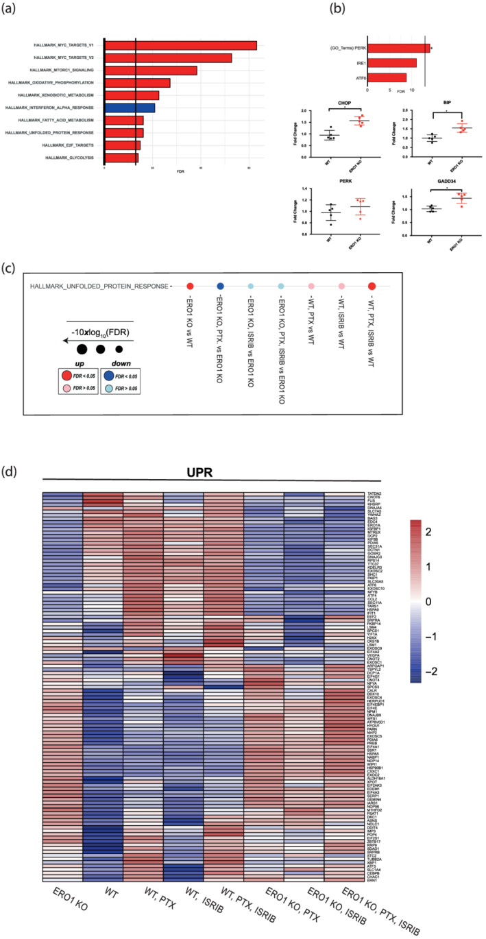
Background and purpose: Endoplasmic reticulum (ER) stress triggers an adaptive response in tumours which fosters cell survival and resilience to stress. Activation of the ER stress response, through its PERK branch, promotes phosphorylation of the α-subunit of the translation initiation factor eIF2, thereby repressing general protein translation and augmenting the translation of ATF4 with the downstream CHOP transcription factor and the protein disulfide oxidase, ERO1-alpha EXPERIMENTAL APPROACH: Here, we show that ISRIB, a small molecule that inhibits the action of phosphorylated eIF2alpha, activating protein translation, synergistically interacts with the genetic deficiency of protein disulfide oxidase ERO1-alpha, enfeebling breast tumour growth and spread.

Key results: ISRIB represses the CHOP signal, but does not inhibit ERO1. Mechanistically, ISRIB increases the ER protein load with a marked perturbing effect on ERO1-deficient triple-negative breast cancer cells, which display impaired proteostasis and have adapted to a low client protein load in hypoxia, and ERO1 deficiency impairs VEGF-dependent angiogenesis. ERO1-deficient triple-negative breast cancer xenografts have an augmented ER stress response and its PERK branch. ISRIB acts synergistically with ERO1 deficiency, inhibiting the growth of triple-negative breast cancer xenografts by impairing proliferation and angiogenesis.

Conclusion and implications: These results demonstrate that ISRIB together with ERO1 deficiency synergistically shatter the PERK-dependent adaptive ER stress response, by restarting protein synthesis in the setting of impaired proteostasis, finally promoting tumour cytotoxicity. Our findings suggest two surprising features in breast tumours: ERO1 is not regulated via CHOP under hypoxic conditions, and ISRIB offers a therapeutic option to efficiently inhibit tumour progression in conditions of impaired proteostasis.

Keywords: ERO1 alpha; ISRIB (integrated stress response inhibitor); PERK pathway; UPR (unfolded protein response); breast cancer; endoplasmic reticulum stress.


Conflict of interest statement

The authors declare no competing interests.

Figures

FIGURE 1
FIGURE 1
ERO1 deficiency promotes proteotoxicity and represses protein translation under hypoxic conditions, which is rescued by ISRIB. (a) Representative immunoblot of detergent‐soluble and ‐insoluble FLAG‐VEGF121 and BIP. Actin was used as a loading control. On the right, dot plots showing the ratio of detergent‐insoluble to detergent‐soluble VEGF121 and BIP. Ratio >1 indicates aggregates, and thus impaired proteostasis (n = 5, unpaired t‐test). (b) Representative immunoblot of newly synthesized, puromycin‐labelled proteins from WT MDAMB231* cells using an anti‐puromycin antibody. Actin was used as a loading control. (c) Puromycin labelling of proteins from ERO1 KO MDAMB231*. (d) Dot plots showing the percentage increase of the puromycin signal between ISRIB‐treated ERO1 KO and WT cells under hypoxic condition (n = 5, unpaired t‐test). (e) Ratio of the viability between ISRIB‐treated ERO1 KO and WT cells under normoxic and hypoxic conditions. Ratio less than 1 indicates impaired viability of ERO1 KO cells (n = 5 for WT and ERO1 KO cells at four different ISRIB concentrations, two‐way ANOVA).
FIGURE 2
FIGURE 2
ISRIB inhibits ATF4 and CHOP signal but not that of ERO1. (a) Quantitative real‐time PCR on cDNA from WT and ERO1 KO MDAMB231* cells (n = 6). (b) ERO1 non‐reducing and reducing western‐blotting. Asterisks mark the different ERO1 bands in non‐reducing conditions; ox. indicates the oxidized ERO1. Ponceau indicates equal protein loading. On the right, dot plot indicating ERO1 levels in arbitrary units (AU) (n = 5). (c) ELISA of VEGF on conditioned media from WT and ERO1 KO MDAMB231* (n = 5). Below, ERO1 western blotting on WT, Het and ERO1 KO MDAMB231*, the asterisk indicates a background band. Actin indicates a loading control. (d) HUVEC migration assay using the conditioned media (CM) from equal numbers of WT and ERO1 KO MDAMB231* cultured in normoxic and hypoxic conditions as a chemoattractant. Differences were calculated by one‐way ANOVA for multiple comparisons.
FIGURE 3
FIGURE 3
ISRIB together with paclitaxel inhibits tumour growth in ERO1 KO MDAMB231* xenografts. (a) Scheme of the pharmacological treatment of mice injected in the mammary fat pad with WT and ERO1 KO MDA‐MB231*. (b) Bioluminescence signals of primary breast tumours from representative mice (n = 10). (c) Bioluminescence signals of ex vivo lungs. (d) Growth curve of breast tumours measured by the caliper and dot plots on a logarithmic scale of the bioluminescence counts of lymph nodes and lung metastases of WT and (e) of ERO1 KO xenografts.
FIGURE 4
FIGURE 4
ISRIB does not restrain tumour angiogenesis but reduces cell viability in necrotic areas. (a) Representative micrographs of CD31 IHC staining in primary breast tumours. Below, relative quantification of CD31+ blood vessels in random fields (n = 5). (b) Representative H&E (haematoxylin & eosin) staining in primary tumours. On the right, H&E staining of necrotic areas. Below, quantification of the number of viable cells in necrotic areas. Differences were calculated by one‐way ANOVA for multiple comparison tests.
FIGURE 5
FIGURE 5
ERO1 KO breast tumours up‐regulate the PERK pathway of the UPR. (a) Bar graphs indicating the top 10 most significantly perturbed gene sets (Hallmark) of ERO1 KO MDAMB231* tumours. Enrichment and their FDR‐adjusted P‐values were computed using a camera (pre‐ranked) and were determined on the Hallmark gene sets collection (MSigDB). The X axis reports the logarithmically transformed FDR value in the form of −10xlog10 (FDR), with a bold intercept (X = 13.01) indicating the FDR threshold of 0.05. Red bars: Up‐regulated; blue bars: Down‐regulated. (b) Bar graphs indicating PERK, IRE1, ATF6 gene sets (GO: Gene ontology gene sets) of ERO1 KO MDAMB231* tumours. PERK pathway was up‐regulated in ERO1 KO MDAMB231* tumours. Below, quantitative real‐time PCR on CHOP (DDIT3), BIP (HSPA5), PERK (EIF2AK3), GADD34 (PPP1R15A) cDNA from WT and ERO1 KO MDAMB231* tumours (N = 5). (c) Dot plots in Hallmark gene sets indicating the up‐regulation and down‐regulation of UPR in WT and ERO1 KO MDAMB231* tumours from mice given the indicated pharmacological treatments (PTX stands for paclitaxel). (d) Heatmap of UPR genes from the Hallmark gene sets collection in WT and ERO1 KO MDAMB231* tumours.
FIGURE 6
FIGURE 6
ERO1/PERK cooperation in breast tumours. Kaplan–Meier plotter depicting relapse‐free survival of breast cancer patients (n = 948) stratified for gene expression levels of ERO1 (a), EIF2AK3 (PERK) (b) and the ratio EIF2AK3/ERO1 (c). In panel (c), the upper (n = 237) and lower quartile (n = 237) of the ratio are represented. Statistical significance was assessed using a log‐rank test.
FIGURE 7
FIGURE 7
ERO1 deficiency in breast tumours up‐regulates PERK and dictates the ISRIB‐mediated cytotoxic effect. ERO1 is a protein disulphide oxidase in the endoplasmic reticulum, whose expression is regulated by CHOP in a variety of ER stress conditions. Previously, we reported that the lack of ERO1 in highly metastatic breast tumours impairs secretion of angiogenic factors, among which VEGFA, and angiogenesis, hence acting on the tumour resilience. In this study, we employed ISRIB, a small molecule which, by reactivating protein translation, enfeebles the adaptive PERK‐mediated mechanism of protein repression. In breast tumour (MDAMB231*) cells under hypoxia, ISRIB inhibits CHOP but has no effect on ERO1 activity, suggesting that under hypoxia CHOP does not regulate ERO1. However, ISRIB is synergistic with ERO1 deficiency in terms of impairment of the tumour burden. Mechanistically, ERO1 deficiency up‐regulates the PERK branch of UPR, repressing protein translation, which renders ISRIB more effective to restrain tumour growth in a context of impaired proteostasis. In ERO1 KO tumours, ISRIB‐dependent reactivation of protein translation together with the impairment of angiogenesis constitutes a double‐hit which weakens tumour resilience to stress.

References

    1. Alexander, S. P. , Fabbro, D. , Kelly, E. , Mathie, A. , Peters, J. A. , Veale, E. L. , Armstrong, J. F. , Faccenda, E. , Harding, S. D. , Pawson, A. J. , & Southan, C. (2021). The concise guide to pharmacology 2021/22: Catalytic receptors. British Journal of Pharmacology, 178(Suppl 1), S264–S312. 10.1111/bph.15541 - DOI - PubMed
    1. Blais, J. D. , Chin, K. T. , Zito, E. , Zhang, Y. , Heldman, N. , Harding, H. P. , Fass, D. , Thorpe, C. , & Ron, D. (2010). A small molecule inhibitor of endoplasmic reticulum oxidation 1 (ERO1) with selectively reversible thiol reactivity. The Journal of Biological Chemistry, 285(27), 20993–21003. doi: M110.126599 [pii]. 10.1074/jbc.M110.126599 - DOI - PMC - PubMed
    1. Claesson‐Welsh, L. , & Welsh, M. (2013). VEGFA and tumour angiogenesis. Journal of Internal Medicine, 273(2), 114–127. 10.1111/joim.12019 - DOI - PubMed
    1. Cruz‐Munoz, W. , Man, S. , Xu, P. , & Kerbel, R. S. (2008). Development of a preclinical model of spontaneous human melanoma central nervous system metastasis. Cancer Research, 68(12), 4500–4505. 10.1158/0008-5472.CAN-08-0041 - DOI - PubMed
    1. Cubillos‐Ruiz, J. R. , Bettigole, S. E. , & Glimcher, L. H. (2017). Tumorigenic and immunosuppressive effects of endoplasmic reticulum stress in cancer. Cell, 168(4), 692–706. doi: S0092‐8674(16)31678‐6 [pii]. 10.1016/j.cell.2016.12.004 - DOI - PMC - PubMed

Publication types

MeSH terms