. 2022 Nov;39(6):903-907.
doi: 10.1111/pde.15094. Epub 2022 Jul 19.

Inflammatory linear verrucous epidermal nevus (ILVEN) encompasses a spectrum of inflammatory mosaic disorders

Affiliations

Inflammatory linear verrucous epidermal nevus (ILVEN) encompasses a spectrum of inflammatory mosaic disorders

Lihi Atzmony et al. Pediatr Dermatol. 2022 Nov.

Abstract

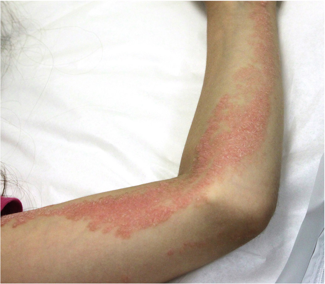
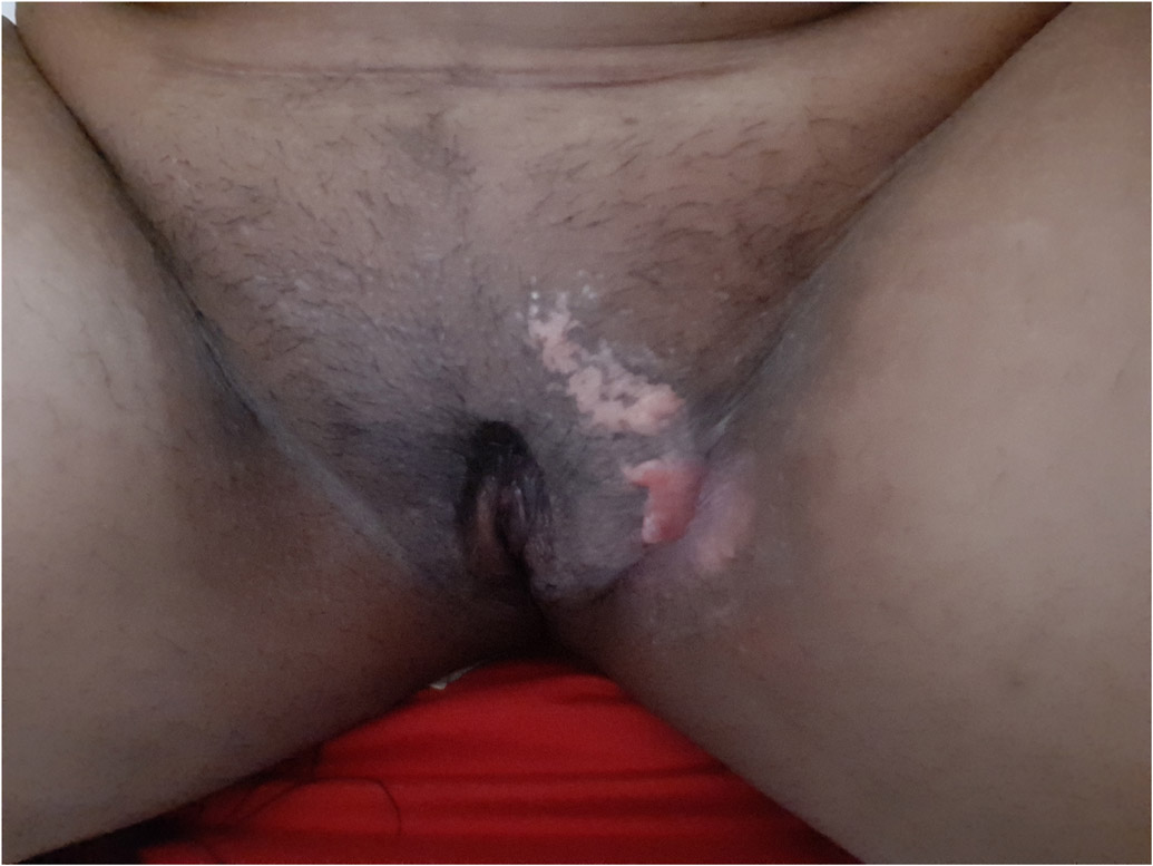
Background: Inflammatory linear verrucous epidermal nevus (ILVEN) is a rare skin disease characterized by pruritic erythematous scaly plaques distributed along the lines of Blaschko. Two cases of ILVEN with CARD14 mutations and one case with a GJA1 mutation have been previously reported.

Objective: To elucidate the genetic cause of a cohort of patients diagnosed based on clinical and histopathological evaluation with ILVEN.

Methods: We recruited patients diagnosed with ILVEN based on clinical and histopathological criteria. Exome sequencing of affected skin with or without blood/saliva was performed and germline and somatic pathogenic variants were identified.

Results: Five patients were enrolled. All had skin lesions from birth or early childhood. Two patients developed psoriasis vulgaris after the diagnosis of ILVEN. The first had a germline heterozygous CARD14 mutation and a post-zygotic hotspot mutation in KRT10. The histopathologic evaluation did not show epidermolytic hyperkeratosis. The second had a post-zygotic hotspot mutation in HRAS. Her ILVEN became itchy once psoriasis developed. One patient was re-diagnosed with linear porokeratosis based on a germline mutation in PMVK and a post-zygotic second-hit mutation. Two patients were re-diagnosed with congenital hemidysplasia with ichthyosiform nevus and limb defect nevus based on germline NSDHL mutations.

Conclusion: ILVEN is a clinical descriptor for a heterogenous group of mosaic inflammatory disorders. Genetic analysis has the potential to more precisely categorize ILVEN and permits pathogenesis-directed therapies in some cases.

Keywords: CHILD syndrome; epidermal nevus; genetic skin diseases; inflammatory linear verrucous epidermal nevus; mosaicism.


Conflict of interest statement

Conflict of Interest

The authors have no conflicts of interest to declare.

Figures

Figure 1.
Figure 1.. Clinical and histologic features of inflammatory linear verrucous epidermal nevus (ILVEN).
a) Patient 1 had a linear verrucous plaque over her right foot and ankle that became itchy once she developed generalized plaque psoriasis. b) Histology demonstrated alternating orthokeratosis and parakeratosis acanthosis and spongiosis. c) Patient 2 had itchy blaschkoid psoriasiform plaques over her left chest and arm. d) Histopathologic evaluation showed psoriasiform epidermal hyperplasia and areas of alternating orthokeratosis and parakeratosis. Some of the keratinocytes showed reticular degeneration with balloon cell morphology. e) Patient 3 presented with extensive whorls of pink plaques, some covered with yellowish crusts. f) Histopathologic evaluation showed alternate parakeratosis overlying hypogranulosis and orthokeratosis, acanthosis and papillomatosis with perivascular and interstitial lymphocytic infiltrate in the superficial dermis. The parakeratosis overlying hypogranulosis corresponds to wide cornoid lamellae. g) Patient4 had a small linear verrucous plaque over her left groin. h) Histopathological evaluation showed psoriasiform hyperplasia with hyperkeratosis and parakeratosis, thin supra-papillary plate and dense lymphohistiocytic infiltrate in the papillary dermis. i) Patient 5 has psoriasiform and warty plaques in a blaschkoid distribution over the left side of her body, including the left groin. Several fingernails had longitudinal leukonychia and the second nail was partially absent. j) Histopathology showed marked parakeratosis and acanthosis and foamy macrophages in the papillary dermis.

References

    1. Morag C, Metzker A. Inflammatory linear verrucous epidermal nevus: report of seven new cases and review of the literature. Pediatr Dermatol. 1985;3(1):15–18. - PubMed
    1. Altman J, Mehregan AH. Inflammatory linear verrucose epidermal nevus. Arch Dermatol. 1971;104(4):385–389. - PubMed
    1. Tiwary A, Mishra D. A unique porokeratotic variant of inflammatory linear verrucous epidermal nevus. Indian J Paediatr Dermatol. 2017;18(3):237–240. doi:10.4103/2319-7250.206088 - DOI
    1. Umegaki-Arao N, Sasaki T, Fujita H, et al. Inflammatory Linear Verrucous Epidermal Nevus with a Postzygotic GJA1 Mutation Is a Mosaic Erythrokeratodermia Variabilis et Progressiva. J Invest Dermatol. 2017;137(4):967–970. doi:10.1016/j.jid.2016.11.016 - DOI - PubMed
    1. Riachi M, Polubothu S, Stadnik P, et al. Molecular Genetic Dissection of Inflammatory Linear Verrucous Epidermal Naevus Leads to Successful Targeted Therapy. J Invest Dermatol. Published online June 8, 2021. doi:10.1016/j.jid.2021.02.765 - DOI - PMC - PubMed