. 2022 Jul 19;12(1):12339.
doi: 10.1038/s41598-022-16504-9.

Differentiation of osteosarcoma from osteomyelitis using microarchitectural analysis on panoramic radiographs

Affiliations

Differentiation of osteosarcoma from osteomyelitis using microarchitectural analysis on panoramic radiographs

Ji-Hun Jung et al. Sci Rep. .

Abstract

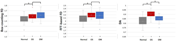
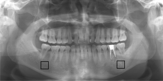
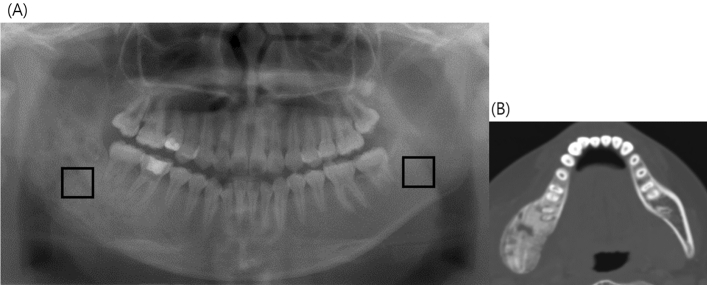
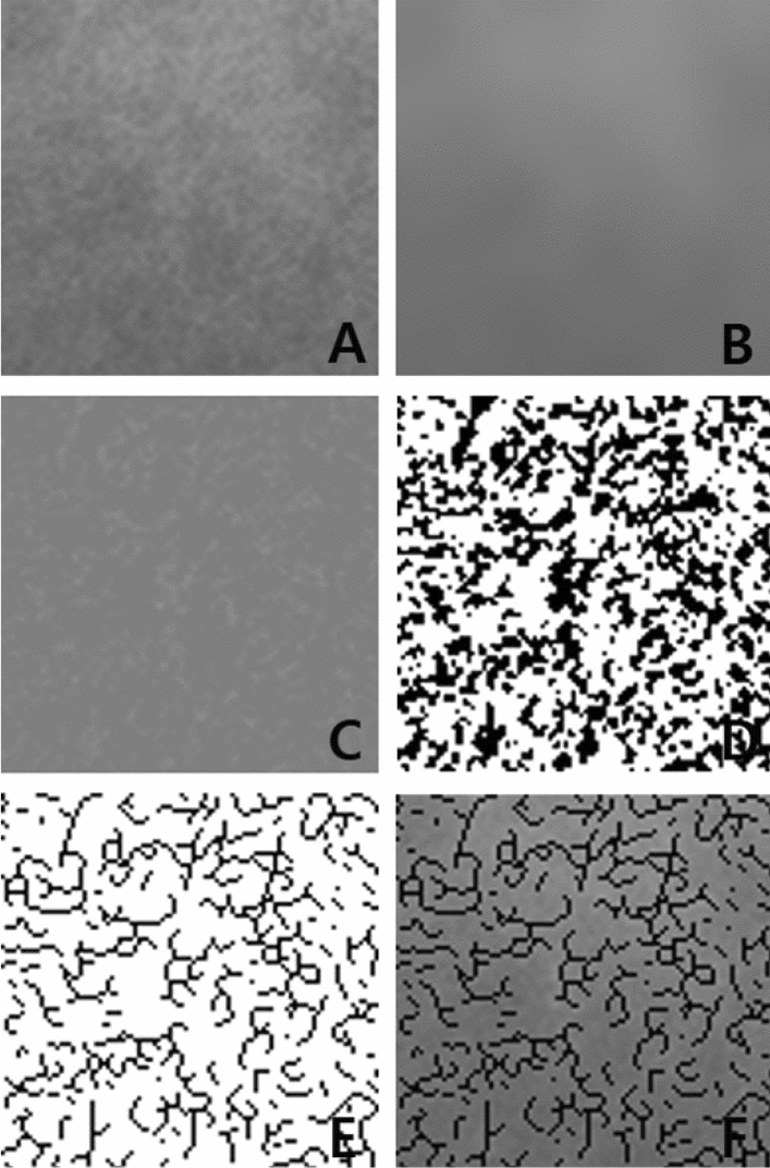
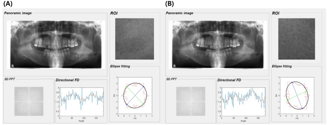
Diagnosing osteosarcoma (OS) is very challenging and OS is often misdiagnosed as osteomyelitis (OM) due to the nonspecificity of its symptoms upon initial presentation. This study investigated the possibility of detecting OS-induced trabecular bone changes on panoramic radiographs and differentiating OS from OM by analyzing fractal dimensions (FDs) and degrees of anisotropy (DAs). Panoramic radiographs of patients with histopathologically proven OS and OM of the jaw were obtained. A total of 23 patients with OS and 40 patients with OM were enrolled. To investigate whether there was a microarchitectural difference between OS lesions and normal trabecular areas in each patient, two regions of interest (ROIs) were located on the CT images. Three microarchitectural parameters (box-counting FD, fast Fourier transform-based FD, and DA) were calculated. For both OS and OM, significant differences were found for all three microarchitectural parameters. Compared to normal trabecular bone, trabecular bone affected by OS and OM became isotropic and more complex. When comparing OS and OM, a statistically significant difference was found only in DA. Trabecular bones affected by OS became more isotropic than those affected by OM. Microarchitectural analysis, especially DA, could be useful for detecting OS-induced trabecular alterations and differentiating OS from OM.


Conflict of interest statement

The authors declare no competing interests.

Figures

Figure 1
Figure 1
Box plot of the degree of anisotropy (DA), fast Fourier transform (FFT)-based fractal dimension (FD), and box-counting FD. A paired t-test was used to compare normal areas and osteosarcoma (OS) lesions. An independent sample t-test was performed to compare OS and osteomyelitis (OM). *Significant difference (P < 0.05).
Figure 2
Figure 2
Panoramic radiograph of a 48-year-old man with osteosarcoma (OS) on the left mandible. Note the location of the two square regions of interest (ROIs): one ROI at the center of the OS on the left mandibular body and the other ROI in the corresponding normal trabecular bone on the right mandibular body.
Figure 3
Figure 3
Panoramic radiograph of a 30-year-old woman with osteomyelitis (OM) on the right mandible. (A) A ROI was located at the center, or the representative area, of the lesion on a panoramic radiograph with reference to the corresponding CT image. The other ROI was placed in the corresponding normal trabecular bone on the left mandibular body. (B) CT image shows the OM lesion of the right posterior mandible more clearly.
Figure 4
Figure 4
Digital analysis of the trabecular bone morphology. (A) A region of interest of the trabecular bone located in the center of the osteosarcoma (OS) area on the left posterior mandible in a patient’s panoramic radiograph in Fig. 1. (B) Result after blurring this region. (C) Result after subtracting (B) from (A) and adding 128 (D) binary versions of the image (C). (E) The trabecular pattern is skeletonized. (F) Addition of images (A) and (E) to visually demonstrate that the skeletonized image corresponds to the original trabeculae.
Figure 5
Figure 5
An example of the fast Fourier transform (FFT)-based method of calculating the fractal dimensions (FDs) and degree of anisotropy (DA) of a panoramic radiograph from patients with osteosarcoma (OS). The DA of the region of interest in the OS (A) on the left mandible was 0.96 and that of the normal trabecular bone (B) on the right mandible was 0.81. OS lesions demonstrated more isotropic trabecular bone structure than the normal area.

References

    1. Ottaviani G, Jaffe N. The epidemiology of osteosarcoma. Cancer Treat. Res. 2009;152:3–13. doi: 10.1007/978-1-4419-0284-9_1,20213383. - DOI - PubMed
    1. Raymond AK, Jaffe N. Osteosarcoma multidisciplinary approach to the management from the pathologist’s perspective. Cancer Treat. Res. 2009;152:63–84. doi: 10.1007/978-1-4419-0284-9_4,20213386. - DOI - PubMed
    1. Meyers PA, et al. Osteogenic sarcoma with clinically detectable metastasis at initial presentation. J. Clin. Oncol. 1993;11:449–453. doi: 10.1200/JCO.1993.11.3.449,8445419. - DOI - PubMed
    1. Kager L, et al. Primary metastatic osteosarcoma: presentation and outcome of patients treated on neoadjuvant cooperative osteosarcoma study group protocols. J. Clin. Oncol. 2003;21:2011–2018. doi: 10.1200/JCO.2003.08.132,12743156. - DOI - PubMed
    1. Mirabello L, Troisi RJ, Savage SA. Osteosarcoma incidence and survival rates from 1973 to 2004: data from the surveillance, epidemiology, and end results program. Cancer. 2009;115:1531–1543. doi: 10.1002/cncr.24121,19197972. - DOI - PMC - PubMed