. 2022 Jul 19;12(1):12302.
doi: 10.1038/s41598-022-16629-x.

SIRT1 upregulation promotes epithelial-mesenchymal transition by inducing senescence escape in endometriosis

Affiliations

SIRT1 upregulation promotes epithelial-mesenchymal transition by inducing senescence escape in endometriosis

Minghua Wang et al. Sci Rep. .

Abstract

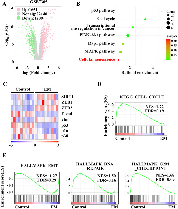
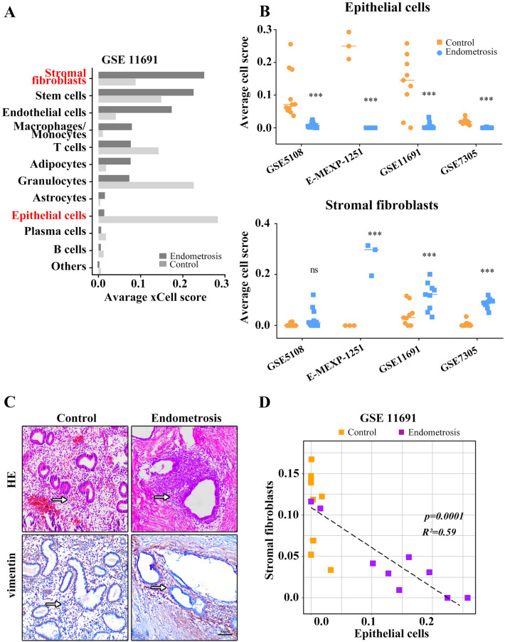
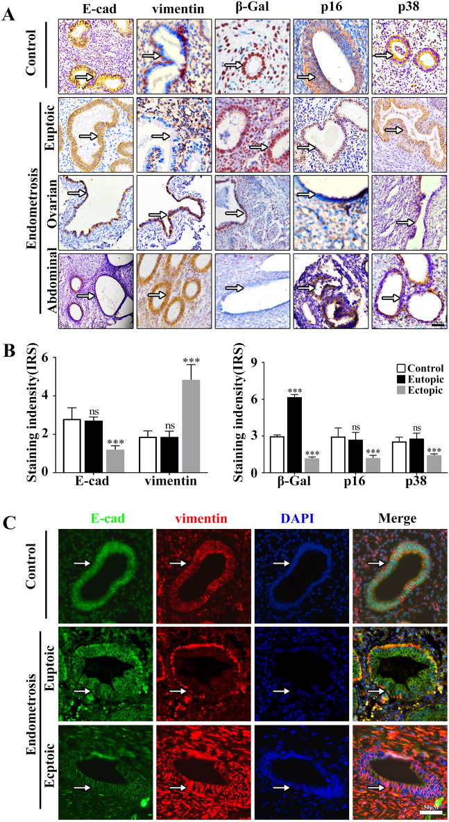
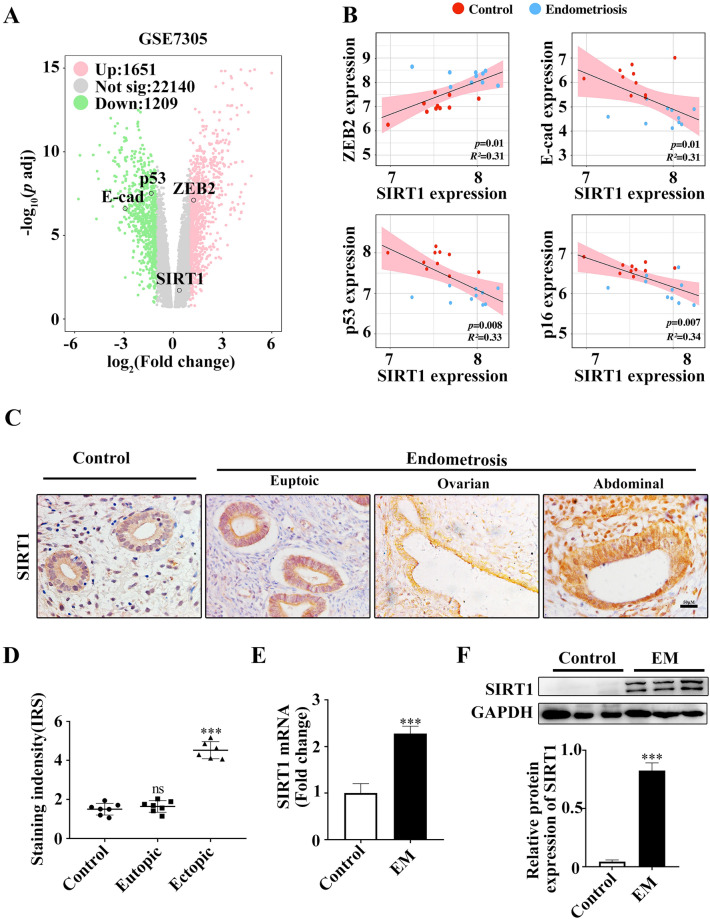
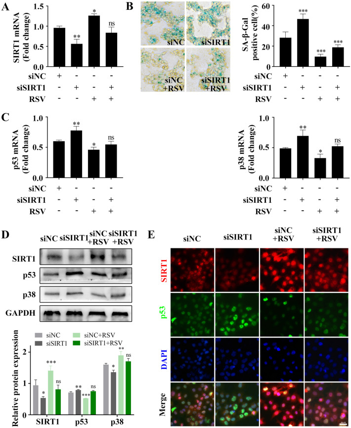
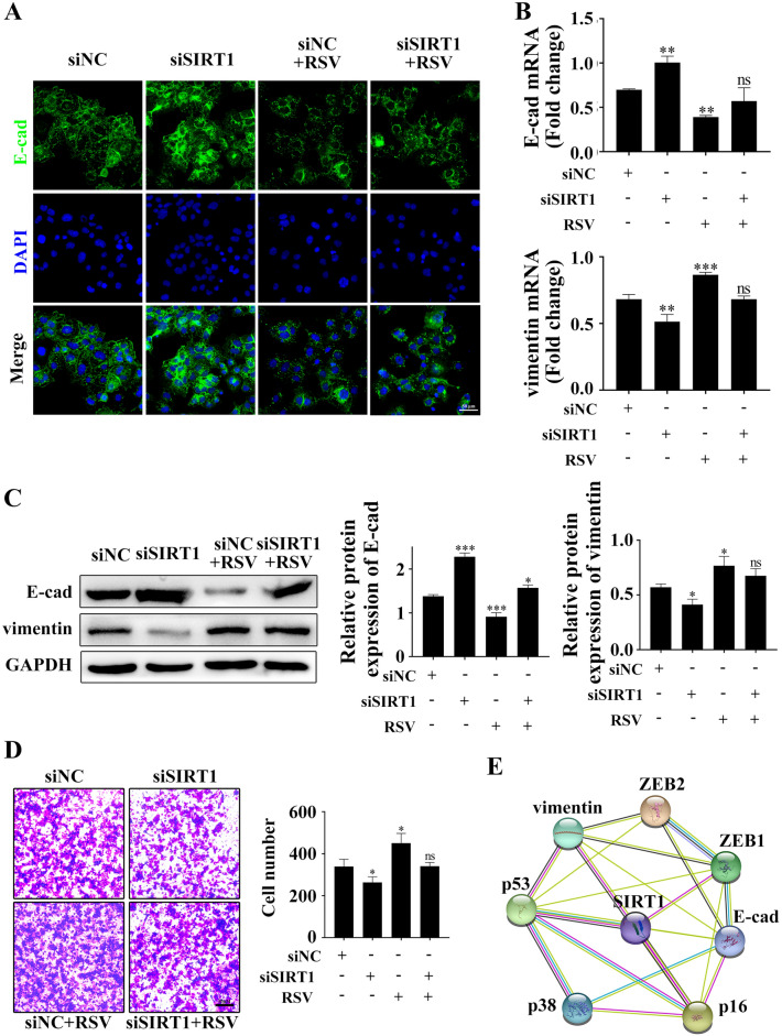
Endometrial epithelial cells carry distinct cancer-associated alterations that may be more susceptible to endometriosis. Mouse models have shown that overexpression of SIRT1 associated with oncogene activation contributes to the pathogenesis of endometriosis, but the underlying reason remains elusive. Here, we used integrated systems biology analysis and found that enrichment of endometrial stromal fibroblasts in endometriosis and their cellular abundance correlated negatively with epithelial cells in clinical specimens. Furthermore, endometrial epithelial cells were characterized by significant overexpression of SIRT1, which is involved in triggering the EMT switch by escaping damage or oncogene-induced induced senescence in clinical specimens and in vitro human cell line models. This observation supports that genetic and epigenetic incident favors endometrial epithelia cells escape from senescence and fuel EMT process in endometriosis, what could be overcome by downregulation of SIRT1.


Conflict of interest statement

The authors declare no competing interests.

Figures

Figure 1
Figure 1
Bioinformatic analysis results support that EMT and senescence are involved in endometriosis. Differentially expressed genes (DEGs) were identified. The red and green indicate unregulated and downregulated DEGs, respectively, in GSE7305 (A). Hallmark pathway enrichment (B) and heatmap of EMT- and senescence-related genes from GSE7305 (C). Endometriosis showing enhanced genes in the “cell cycle” pathway (D). GSEA revealed enrichment of endometriosis genes in “EMT”, “DNA repair” and “G2/M checkpoint” (E).
Figure 2
Figure 2
Relative expression of EMT- and senescence-related genes Expression as shown by qRT-PCR and western blotting, respectively (A,B). Anti-GAPDH antibody was used as loading control. Greyscale values of the bands were determined by Quantity One software. *p < 0.05, **p < 0.01, ***p < 0.001.
Figure 3
Figure 3
Endometrial stromal fibroblasts were enriched in endometriosis compared to other cell types. Computational biology analysis revealed the total content of different cells in the tissues of the control and endometriosis (GSE11691, n = 10) (A). Endometrial stromal fibroblast and epithelial cell abundance scores in tissues of the control and endometriosis, as calculated by xCell (B). HE staining and vimentin immunohistological staining showing stromal fibroblasts and epithelial cells (C). Abundance score correlation analysis for stromal fibroblasts with epithelial cells in tissues of the control and endometriosis (D). Scale bars = 50 µm in (C). ***p < 0.001.
Figure 4
Figure 4
EMT- and senescence-related protein expression in epithelial cells of endometriosis. Immunostaining for E-cad, vimentin, β-gal, p16 and p38 (A). The intensity of the immunohistological staining was given an IRS score as the mean ± SEM. The results show that E-cad and vimentin expression was stronger, but β-gal, p16 and p38 expression was weaker in epithelial cells in endometriosis tissues than in the controls (B). E-cad and vimentin protein levels were examined by immunofluorescence staining (C). Scale bars = 50 µm in (A,C). ***p < 0.001.
Figure 5
Figure 5
The expression of SIRT1 was unregulated in endometriosis. A volcano plot shows the high expression of SIRT1 and ZEB2 and the low expression of E-cad and p53 in endometriosis (A). Correlation analysis of SIRT1 with ZEB2, E-cad, p53 and p16 in endometriosis (B). Representative images of SIRT1 expression were detected by IHC in control and endometriotic endometria (C). Dot plot of the IRS scores. The differences were analysed by Student’s t-test. ***p < 0.001 (D). qPCR (E) and western blots (F) were used to detect the SIRT1 expression levels in the control and endometriotic endometrium. E-cad E-cadherin, EM endometriosis. Scale bars = 50 µm in (C).
Figure 6
Figure 6
Effect of RSV on the senescence of Ishikawa cells. qPCR analysis of SIRT1 mRNA levels in Ishikawa cells first transfected with siNC or siSIRT1 and then treated with or without RSV for 24 h (A). Representative SaβG staining in parallel Ishikawa cell cultures (B). qPCR analysis of p53 and p38 mRNA levels in Ishikawa cells first transfected with siNC or siSIRT1 and then treated with or without RSV for 24 h (C). Representative western blot and quantification of senescence-related genes from parallel cell cultures (D). p53 and SIRT1 protein levels were examined by immunofluorescence staining (E). RSV resveratrol. Scale bars = 50 µm in (E). *p < 0.05, **p < 0.01, ***p < 0.001.
Figure 7
Figure 7
Effect of RSV on the EMT of Ishikawa cells. Ishikawa cells were treated with siNC or siSIRT1 with or without RSV, and then E-cad protein levels were examined by immunofluorescence staining (A). E-cad and vimentin mRNA or protein levels were examined by qPCR (B) and western blotting (C). Matrigel invasion assays to determine the invasive properties of the cells from the parallel cell cultures (D). The bioinformatics analysis depicting the correlation between SIRT1, ECM and senescence was mapped with the STRING database to construct a PPI network (E). Scale bars = 50 µm in (A). RSV resveratrol, E-cad E-cadherin. *p < 0.05, **p < 0.01, ***p < 0.001.

References

    1. Gordts S, Koninckx P, Brosens I. Pathogenesis of deep endometriosis. Fertil. Steril. 2017;108:872–885. doi: 10.1016/j.fertnstert.2017.08.036. - DOI - PubMed
    1. Guo SW. Cancer-associated mutations in endometriosis: Shedding light on the pathogenesis and pathophysiology. Hum. Reprod. Update. 2020;26:423–449. doi: 10.1093/humupd/dmz047. - DOI - PubMed
    1. Guo SW. Fibrogenesis resulting from cyclic bleeding: The Holy Grail of the natural history of ectopic endometrium. Hum. Reprod. 2018;33:353–356. doi: 10.1093/humrep/dey015. - DOI - PubMed
    1. Chen M, et al. Bioinformatic analysis reveals the importance of epithelial-mesenchymal transition in the development of endometriosis. Sci. Rep. 2020;10:8442. doi: 10.1038/s41598-020-65606-9. - DOI - PMC - PubMed
    1. Poli-Neto OB, Meola J, Rosa-E-Silva JC, Tiezzi D. Transcriptome meta-analysis reveals differences of immune profile between eutopic endometrium from stage I-II and III-IV endometriosis independently of hormonal milieu. Sci. Rep. 2020;10:313. doi: 10.1038/s41598-019-57207-y. - DOI - PMC - PubMed

Publication types