Review
. 2022 Jul 19;17(1):279.
doi: 10.1186/s13023-022-02421-7.

A recurrent single-amino acid deletion (p.Glu500del) in the head domain of ß-cardiac myosin in two unrelated boys presenting with polyhydramnios, congenital axial stiffness and skeletal myopathy

Affiliations
Review

A recurrent single-amino acid deletion (p.Glu500del) in the head domain of ß-cardiac myosin in two unrelated boys presenting with polyhydramnios, congenital axial stiffness and skeletal myopathy

Ingrid Bader et al. Orphanet J Rare Dis. .

Abstract

Background: Alterations in the MYH7 gene can cause cardiac and skeletal myopathies. MYH7-related skeletal myopathies are extremely rare, and the vast majority of causal variants in the MYH7 gene are predicted to alter the rod domain of the of ß-cardiac myosin molecule, resulting in distal muscle weakness as the predominant manifestation. Here we describe two unrelated patients harboring an in-frame deletion in the MYH7 gene that is predicted to result in deletion of a single amino acid (p.Glu500del) in the head domain of ß-cardiac myosin. Both patients display an unusual skeletal myopathy phenotype with congenital axial stiffness and muscular hypertonus, but no cardiac involvement.

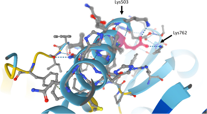
Results: Clinical data, MRI results and histopathological data were collected retrospectively in two unrelated boys (9 and 3.5 years old). Exome sequencing uncovered the same 3-bp in-frame deletion in exon 15 (c.1498_1500delGAG) of the MYH7 gene of both patients, a mutation which deletes a highly conserved glutamate residue (p.Glu500del) in the relay loop of the head domain of the ß-cardiac myosin heavy chain. The mutation occurred de novo in one patient, whereas mosaicism was detected in blood of the father of the second patient. Both boys presented with an unusual phenotype of prenatal polyhydramnios, congenital axial stiffness and muscular hypertonus. In one patient the phenotype evolved into an axial/proximal skeletal myopathy without distal involvement or cardiomyopathy, whereas the other patient exhibited predominantly stiffness and respiratory involvement. We review and compare all patients described in the literature who possess a variant predicted to alter the p.Glu500 residue in the ß-cardiac myosin head domain, and we provide in-silico analyses of potential effects on polypeptide function.

Conclusion: The data presented here expand the phenotypic spectrum of mutations in the MYH7 gene and have implications for future diagnostics and therapeutic approaches.

Keywords: Actin-binding domain; Exome-analysis; MYH7; Myopathy; ß-cardiac myosin heavy chain.


Conflict of interest statement

The authors declare they have no competing interests.

Figures

Fig. 1
Fig. 1
MRI findings of patient 1. Top row, T2 w MRI. (B + D) Sagittal (B) and frontal (D) view of the neck muscles of patient 1 at the age of 7 years. Neck muscles and paravertebral muscles are degenerated with compensatory hyperlordosis of the cervical spine compared to (A + C) age-matched healthy controls. Second row shows the difference between right and left T2 w MRI at the age of 7 years: (E + F) frontal view of right + left shoulder showing symmetrical fatty degeneration of the supraspinatus muscle (S.m.) and involvement of the infraspinatus muscles (I.m.) compared to normal signal for deltoid muscles (D.m. in red). Bottom row shows the difference between right and left T1 w MRI at the age of 9 years: (G) Frontal view of thigh with obturator internus (O.a.m.) and adductor brevis (A.b.m) muscles replaced by fat tissue, adductor longus (A.l.m.) and adductor magnus (A.m.m.) muscles fatty degenerated. (H) A proximal axial view of the hip muscles: with the left gluteus maximus muscle (G.m.m.) showing slight signs of fatty infiltration, the right side appears not to be affected. (I + J) A distal axial view of the thigh muscle showing very slight involvement of the left biceps femoris, sartorius and semimembranosus muscles (circled in yellow) and no obvious involvement of the right thigh
Fig. 2
Fig. 2
MYH7 mutation p.Glu500del. (A + B) Electropherograms from Sanger sequencing of DNA from blood of A patient 1 (top row) and his parents show the 3-bp in-frame deletion in exon 15: MYH7 (NM_000257.3) c.1498_1500delGAG p.(Glu500del) heterozygous, de novo. B Patient 2 (top row) and his parents: the 3-bp in-frame deletion in exon 15 is present in patient 2 (top row) and also in a fraction of approx. 38% in DNA from blood of the father, as estimated from the area under the curve of the three bases after the frameshift in the electropherogram (bottom row). C Graphic representation of the 1935-amino-acid human beta myosin heavy chain polypeptide and location of p.Glu500del. Domains adapted from Colgrave 2014 and derived from the reference-sequence NM_000248.2: head domain (aa 1–847, blue) including the actin-binding domain (aa 655-677 and 757–771), relay-loop (aa 490–513, green), neck-domain (aa 848–1216, orange), rod-domain (aa 1217–1935, red). D MSA of Myh7 orthologs: Homo sapiens (NP_000248.2), Mus musculus (NP_542766.1); chicken, Gallus gallus (NM_001001302); Xenopus laevis (NM_001091682); Danio rerio (NP_001070932.2); scallop, Argopecten irradians (X55714.1); Caenorhabditis elegans (NP_724006.1), Drosophila melanogaster (NP_724006.1), Arabidopsis thaliana (NP_188630.1), Saccharomyces cerevisiae (NP_014971.1), Dictyostelium discoideum (XP_645195.1), Saccharomyces cerevisiae (NP_014971.1). The deleted amino acid position p.Glu500del is indicated by a box. The conserved relay loop (aa 490–513) is indicated by a double-headed arrow
Fig. 3
Fig. 3
AD Patient 1: Stained muscle biopsy from adductor muscle at the age of 18 months. A Note increased variability of muscle fiber diameters, endomysial fibrosis (open arrows), endomysial fat cells (asterisks) and muscle fibers with internal myonuclei (arrows). H and E. B Staining for nicotinamide adenine dinucleotide tetrazolium reductase (NADH-TR): note a-reactive, irregularly shaped sarcoplasmatic structures (arrows) displaying a reducing body. C Immunohistochemistry using an antibody against type-I myosin displays type-I fiber predominance. D Electron microscopy: electrodense, granulofilamentous sarcoplasmic structures resembling a “reducing body” (arrows). (E + F) Patient 2: Stained muscle biopsy (E) H and E: Increased variability of muscle fiber diameters and proliferation of endomysial connective tissue (arrows). (F) Cytochrome-c-oxidase: uniformly stained muscle fibers compatible with type-I fiber predominance
Fig. 4
Fig. 4
Intramolecular interactions involving p.Glu500, as predicted by AlphaFold. Close-up on the alpha-helix of the relay loop. p.Glu500 (colored in pink) is situated close to one end of the helix and is expected to engage in electrostatic interactions (dashed blue lines) with Lysine503 and Lysine762. Deletion of p.Glu500 may abolish these interactions and dislocate the positively charged amino group of Lysine503

References

    1. Nag S, Trivedi DV, Sarkar SS, et al. The myosin mesa and the basis of hypercontractility caused by hypertrophic cardiomyopathy mutations. Nat Struct Mol Biol. 2017;24:525–533. doi: 10.1038/nsmb.3408. - DOI - PMC - PubMed
    1. Beecroft SJ, Van De Locht M, De Winter JM, et al. Recessive MYH7-related myopathy in two families. Neuromuscul Disord. 2019;29:456–467. doi: 10.1016/j.nmd.2019.04.002. - DOI - PubMed
    1. Colegrave M, Peckham M. Structural implications of beta-cardiac myosin heavy chain mutations in human disease. Anat Rec (Hoboken) 2014;297:1670–1680. doi: 10.1002/ar.22973. - DOI - PubMed
    1. Mamelona J, Filice L, Oussedik Y, et al. A novel missense mutation in the MYH7 gene causes an uncharacteristic phenotype of myosin storage myopathy: a case report. BMC Med Genet. 2019;20:78. doi: 10.1186/s12881-019-0804-0. - DOI - PMC - PubMed
    1. Dabaj I, Carlier RY, Gomez-Andres D, et al. Clinical and imaging hallmarks of the MYH7-related myopathy with severe axial involvement. Muscle Nerve. 2018;58:224–234. doi: 10.1002/mus.26137. - DOI - PubMed

Publication types