

The early association of water irrigation with negative pressure wound therapy does not more efficiently reduce the depth of the alkali infiltration progress into the burn
- PMID: 35854477
- PMCID: PMC9885477
- DOI: 10.1111/iwj.13883
The early association of water irrigation with negative pressure wound therapy does not more efficiently reduce the depth of the alkali infiltration progress into the burn
Abstract
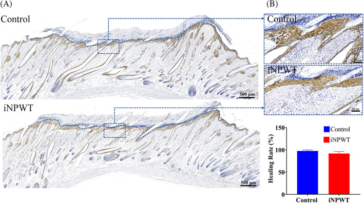
Water irrigation is an efficacious decontaminating method for dermis exposures to corrosive agents and hence has been widely applied to treat especially alkali burns. Nevertheless, once alkali has infiltrated the deep subcutaneous tissue, washing the tissue surface with water irrigation does not attenuate the damage progress. Therefore, significant efforts have been devoted to promising strategies aimed at removing the deeply infiltrated lye. According to a recent report, the negative pressure wound therapy (NPWT) reduces the pH value of the exudate from alkali-provoked burns thus accelerating wound healing. However, it remains to be ascertained whether or not NPWT coupled with water irrigation, that is, iNPWT, more effectively hinders the alkali injury deepening. In this study, we compared the effectiveness of an early application of water irrigation with or without NPWT in preventing the progressive deepening of the alkali burn in an animal model. Our histological examination results showed no appreciable difference in tissue injury depth, dermal retention, inflammatory cell infiltration, re-epithelization, and cellular function between iNPWT and water irrigation alone treatments. Thus, our results prove that the more expensive NPWT coupled with water irrigation does not more effectively hinder the alkali's injury deepening. Hence, iNPWT use should be more cautious in clinical practice.
Keywords: NPWT; alkali burn; water irrigation; wound healing.
© 2022 The Authors. International Wound Journal published by Medicalhelplines.com Inc (3M) and John Wiley & Sons Ltd.
Conflict of interest statement
The authors declare that there is no conflict of interest.
Figures


References
-
- Hall AH, Mathieu L, Maibach HI. Acute chemical skin injuries in the United States: a review. Crit Rev Toxicol. 2018;48(7):540‐554. - PubMed
-
- Li W, Wu X, Gao C. Ten‐year epidemiological study of chemical burns in Jinshan, Shanghai, Pr China. Burns. 2013;39(7):1468‐1473. - PubMed
-
- Yano K, Hata Y, Matsuka K, Ito O, Matsuda H. Experimental study on alkaline skin injuries—periodic changes in subcutaneous tissue pH and the effects exerted by washing. Burns. 1993;19(4):320‐323. - PubMed
-
- Robinson EP, Chhabra AB. Hand chemical burns. J Hand Surg Am. 2015;40(3):605‐612. quiz 13. - PubMed
-
- Leonard LG, Scheulen JJ, Munster AM. Chemical burns: effect of prompt first aid. J Trauma. 1982;22(5):420‐423. - PubMed
MeSH terms
Substances
Grants and funding
- 2020A1515010613/Guangdong Basic and Applied Basic Research Foundation
- 2021A1515220176/Guangdong Basic and Applied Basic Research Foundation
- A2021077/Guangdong Medical Science and Technology Research Foundation
- 82172214/National Natural Science Foundation of China
- 82072180/National Natural Science Foundation of China
LinkOut - more resources
Full Text Sources
