

Pineal region ganglioglioma: A neoplasm with a bimodal age distribution
- PMID: 35855114
- PMCID: PMC9282777
- DOI: 10.25259/SNI_443_2022
Pineal region ganglioglioma: A neoplasm with a bimodal age distribution
Abstract
Background: Gangliogliomas arise very rarely in the pineal region, where their natural histories and pathologic features are poorly understood.
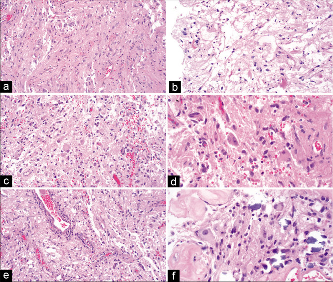
Case description: In this report, we describe a 36-year-old woman who presented with a seizure followed by worsening headache, dizziness, confusion, and intermittent left facial numbness over the next few weeks. A head CT scan showed a partially calcified pineal region mass with hydrocephalus. After an endoscopic third ventriculostomy, the patient underwent a resection of the tumor that contained dysplastic ganglion cells and piloid glial cells. Molecular profiling of this CNS WHO Grade 1 ganglioglioma revealed polysomies of chromosomes 7 and 9, and a BUB1 variant of uncertain significance, without known MAP kinase pathway alterations. From a review of the literature, we found two distinct age distributions for pineal ganglioglioma, with modes at 1 and 36 years of age.
Conclusion: Although very rare, this tumor should be considered in the differential diagnosis of pineal region tumors in children and young adults.
Keywords: Chromosome 7 polysomy; Epiphysis; Glioneuronal; Targeted next-generation sequencing; Tumor.
Copyright: © 2022 Surgical Neurology International.
Conflict of interest statement
There are no conflicts of interest.
Figures


Similar articles
-
Malignant papillary glioneuronal tumor of the pineal gland: Case presentation and literature review of a distinct entity.Am J Case Rep. 2013 May 22;14:164-8. doi: 10.12659/AJCR.883919. Print 2013. Am J Case Rep. 2013. PMID: 23826458 Free PMC article.
-
Pineal ganglioglioma in a young girl: a case report and review of the literature.J La State Med Soc. 2004 Nov-Dec;156(6):316-8. J La State Med Soc. 2004. PMID: 15688672 Review.
-
High-Grade Temporal Ganglioglioma in an Older Adult Woman.Cureus. 2023 Sep 24;15(9):e45862. doi: 10.7759/cureus.45862. eCollection 2023 Sep. Cureus. 2023. PMID: 37881386 Free PMC article.
-
Reclassification of pineal tumor as high-grade astrocytoma with piloid features through methylation profiling: illustrative case.J Neurosurg Case Lessons. 2025 May 5;9(18):CASE24778. doi: 10.3171/CASE24778. Print 2025 May 5. J Neurosurg Case Lessons. 2025. PMID: 40324324 Free PMC article.
-
[Ganglioglioma of the pineal region: case report].Arq Neuropsiquiatr. 2001 Sep;59(3-A):599-604. Arq Neuropsiquiatr. 2001. PMID: 11588644 Review. Portuguese.
References
-
- Burres KP, Hamilton RD. Pineal apoplexy. Neurosurgery. 1979;4:264–8. - PubMed
-
- Camins MB, Schlesinger EB. Treatment of tumours of the posterior part of the third ventricle and the pineal region: A long term follow-up. Acta Neurochir (Wien) 1978;40:131–43. - PubMed
-
- Chang YL, Lin SZ, Chiang YH, Liu MY, Lee WH. Pineal ganglioglioma with premature thelarche. Report of a case and review of the literature. Childs Nerv Syst. 1996;12:103–6. - PubMed
-
- Da Costa MD, Centeno R, Neto M, Dória-Netto H, Filho J, de Araujo Paz D, et al. Anaplastic ganglioglioma of the pineal region-a case report. Arq Bras Neurocir. 2015;35:253–6.
Publication types
LinkOut - more resources
Full Text Sources