

Intramedullary cervical spinal cord and cerebellar hemangioblastoma: A case report
- PMID: 35855144
- PMCID: PMC9282796
- DOI: 10.25259/SNI_525_2022
Intramedullary cervical spinal cord and cerebellar hemangioblastoma: A case report
Abstract
Background: Hemangioblastomas are benign tumors that develop in the central nervous system. They represent 1.5-2.5% of all intracranial tumors, and about 2-15% of all spinal cord tumors. They are highly associated with von Hippel-Lindau disease.
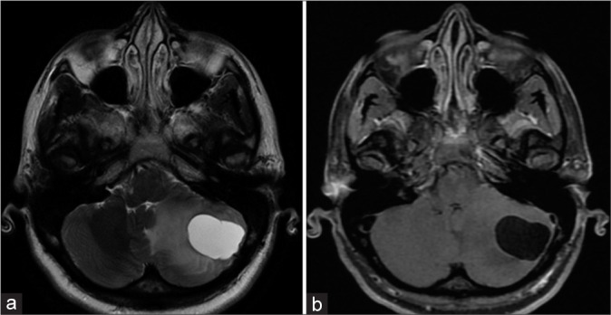
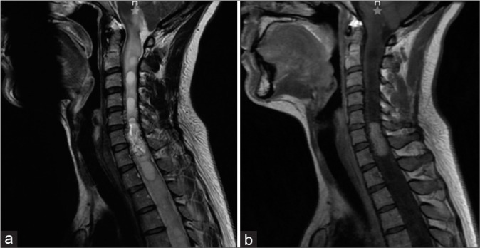
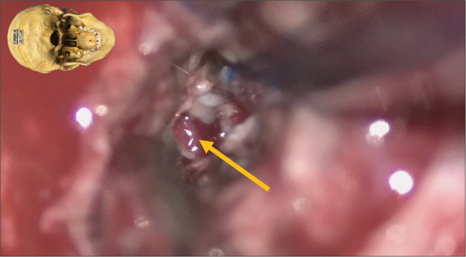
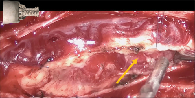
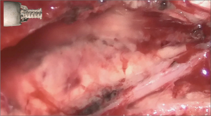
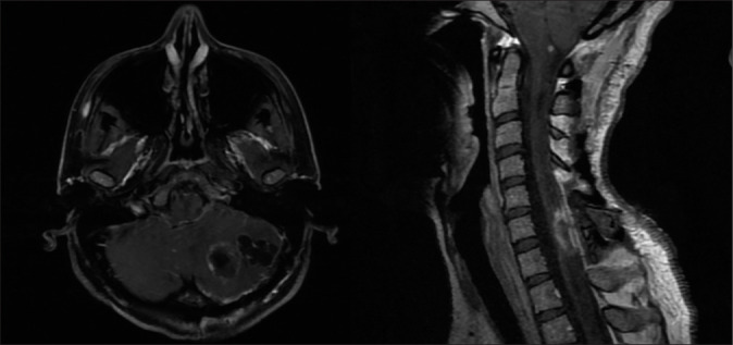
Case description: A 36-year-old female presented with a 4-year history of progressive right upper extremity distal weakness and cervical pain. The magnetic resonance imaging demonstrated a homogeneously, contrast enhancing intradural/intramedullary tumor at C6-C7 with perilesional edema and a syrinx accompanied by a cerebellar cyst with a mural nodule. Surgery included excision of the spinal lesion and decompression and excision of the cerebellar cyst and mural nodule (i.e., median suboccipital craniectomy and cervical C5-C7 laminectomy).
Conclusion: Surgery is the gold standard treatment for symptomatic hemangioblastomas, and surgical approaches should minimize risk.
Keywords: Hemangioblastoma; Neurosurgery; Spine; Spine surgery; von Hippel-Lindau.
Copyright: © 2022 Surgical Neurology International.
Conflict of interest statement
There are no conflicts of interest.
Figures
Similar articles
-
Intradural extramedullary hemangioblastoma of the thoracic cord: A case report.Surg Neurol Int. 2021 Mar 30;12:126. doi: 10.25259/SNI_795_2020. eCollection 2021. Surg Neurol Int. 2021. PMID: 33880231 Free PMC article.
-
Hemorrhagic intramedullary hemangioblastoma of the cervical spinal cord presenting with acute-onset quadriparesis: case report and review of the literature.J Spinal Cord Med. 2014 Nov;37(6):791-4. doi: 10.1179/2045772314Y.0000000210. Epub 2014 Jul 16. J Spinal Cord Med. 2014. PMID: 25029412 Free PMC article. Review.
-
The natural history of hemangioblastomas of the central nervous system in patients with von Hippel-Lindau disease.J Neurosurg. 2003 Jan;98(1):82-94. doi: 10.3171/jns.2003.98.1.0082. J Neurosurg. 2003. PMID: 12546356
-
Intradural Extramedullary Hemangioblastoma of the Cervical Spine: Case Report and Literature Review.Cureus. 2022 May 18;14(5):e25125. doi: 10.7759/cureus.25125. eCollection 2022 May. Cureus. 2022. PMID: 35733499 Free PMC article.
-
Primary Intradural Extramedullary Sporadic Spinal Hemangioblastomas: Case Report and Systematic Review.World Neurosurg. 2021 Aug;152:84-94. doi: 10.1016/j.wneu.2021.05.105. Epub 2021 Jun 1. World Neurosurg. 2021. PMID: 34087464
References
-
- Amano T, Tokunaga S, Shono T, Mizoguchi M, Matsumoto K, Yoshida F, et al. Cerebellar haemangioblastoma manifesting as hearing disturbance. Neurol Med Chir (Tokyo) 2009;49:418–20. - PubMed
-
- Kanno H, Yamamoto I, Nishikawa R, Matsutani M, Wakabayashi T, Yoshida J, et al. Spinal cord hemangioblastomas in von Hippel-Lindau disease. Spinal Cord. 2009;47:447–52. - PubMed
-
- Kuharic M, Jankovic D, Splavski B, Boop FA, Arnautovic KI. Hemangioblastomas of the posterior cranial fossa in adults: Demographics, clinical, morphologic, pathologic, surgical features, and outcomes. A systematic review. World Neurosurg. 2018;110:e1049–62. - PubMed
-
- Mandigo CE, Ogden AT, Angevine PD, McCormick PC. Operative management of spinal hemangioblastoma. Neurosurgery. 2009;65:1166–77. - PubMed
Publication types
LinkOut - more resources
Full Text Sources
Miscellaneous