

Superior sagittal sinus dural arteriovenous fistula with changes in angiographic findings associated with contiguous parasagittal meningioma: A case report
- PMID: 35855145
- PMCID: PMC9282788
- DOI: 10.25259/SNI_95_2022
Superior sagittal sinus dural arteriovenous fistula with changes in angiographic findings associated with contiguous parasagittal meningioma: A case report
Abstract
Background: Meningioma and dural arteriovenous fistula (dAVF) located at the same site are rare. The present case demonstrated the transformation of tumor feeding vessels into the pial feeder of the dAVF over time, which may help to elucidate the pathogenesis of tumor-associated dAVF.
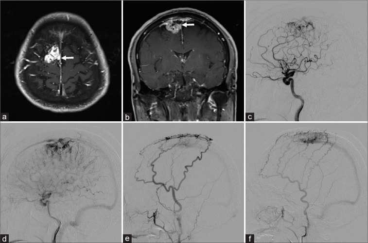
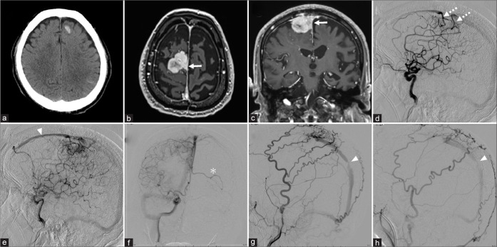
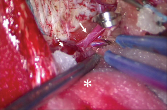
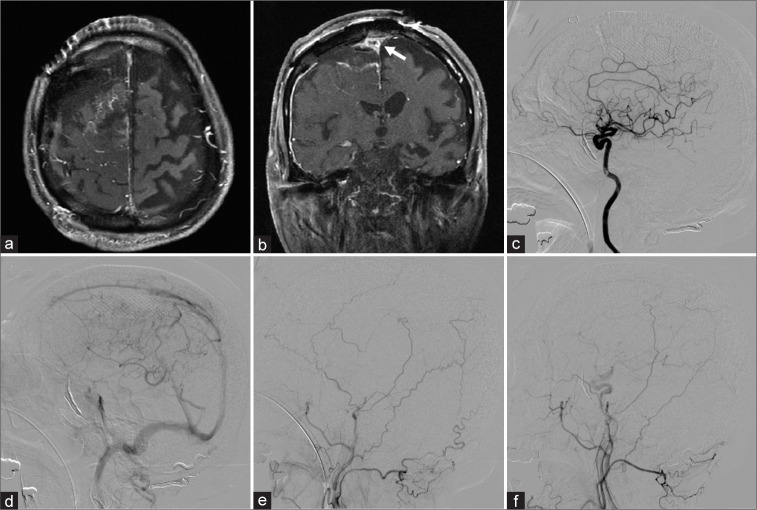
Case description: A 71-year-old man presented with convulsion. Magnetic resonance (MR) imaging showed a right parasagittal sinus meningioma invading the superior sagittal sinus (SSS). Bilateral external carotid angiography showed dAVF at the SSS, near the site of tumor invasion. The right internal carotid angiography showed tumor staining from the anterior cerebral artery with intra-tumor arteriovenous shunting, with stagnation of tumor blood flow, suggesting impairment of perfusion to the SSS. Four years after the initial diagnosis, the patient was admitted to hospital with status epilepticus, and MR imaging showed an enlarged tumor. Carotid angiography revealed transformation of the tumor feeders to the pial feeder of the dAVF. The findings of shunting to the SSS had intensified, and stenosis had occurred in the posterior third of the SSS. The venous return showed retrograde flow anteriorly to the SSS. The patient underwent endovascular embolization and tumor resection. The shunt had disappeared.
Conclusion: This report supports the proposal that impaired venous return is an important factor in the shunt occurrence of dAVF. Neurosurgeons should consider that cases of meningioma invading the venous sinuses may be complicated by dAVF and changes may occur over time.
Keywords: Dural arteriovenous fistula; Meningioma; Venous pressure.
Copyright: © 2022 Surgical Neurology International.
Conflict of interest statement
There are no conflicts of interest.
Figures
References
-
- Ahn JY, Lee BH, Cho YJ, Joo JY, Lee KS. Dural arteriovenous fistula associated with meningioma: Spontaneous disappearance after tumor removal. Neurol Med Chir (Tokyo) 2003;43:308–11. - PubMed
-
- Arnautovic KI, Al-Mefty O, Angtuaco E, Phares LJ. Dural arteriovenous malformations of the transverse/sigmoid sinus acquired from dominant sinus occlusion by a tumor: Report of two cases. Neurosurgery. 1998;42:383–8. - PubMed
-
- Cognard C, Gobin YP, Pierot L, Bailly AL, Houdart E, Casasco A, et al. Cerebral dural arteriovenous fistulas: Clinical and angiographic correlation with a revised classification of venous drainage. Radiology. 1995;194:671–80. - PubMed
-
- Enatsu R, Asahi M, Matsumoto M, Hirai O. Meningioma-related dural arteriovenous fistula fed via a vascular tumor bed: A case report and literature review. Clin Neurol Neurosurg. 2012;114:1010–3. - PubMed
Publication types
LinkOut - more resources
Full Text Sources