

Intradural, extramedullary hemangioblastoma at the level of the conus medullaris: illustrative case
- PMID: 35855219
- PMCID: PMC9245781
- DOI: 10.3171/CASE2145
Intradural, extramedullary hemangioblastoma at the level of the conus medullaris: illustrative case
Abstract
Background: Hemangioblastomas are rare, slow-growing, and highly vascularized tumors that typically occur in the cerebellum and spinal cord. The cervical and thoracic regions are the most common spinal sites, and the tumors are usually intramedullary.
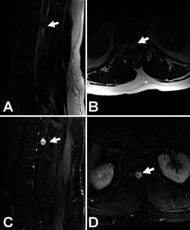
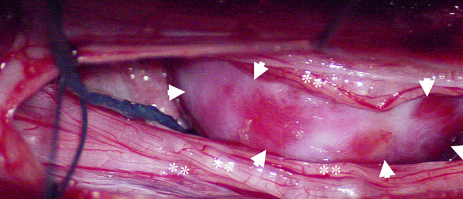
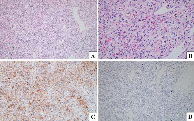
Observations: The authors report the case of a man whose chest computed tomography performed for managing coronavirus disease 2019 incidentally revealed an enhancing area in the spinal canal at T12-L1. The patient reported low back pain as well as leg numbness and tingling. Magnetic resonance imaging of the lumbar region with and without gadolinium contrast demonstrated an intradural, extramedullary lesion with displacement of the conus medullaris. The differential diagnosis included a schwannoma and myxopapillary ependymoma. Bilateral T12-L1 laminectomies were performed with resection of the mass. The general pathologist rendered the frozen section diagnosis of a spindle cell neoplasm, suggesting the differential diagnosis of schwannoma or myxopapillary ependymoma. Immunohistochemistry was positive for inhibin, GFAP, reticulin, CD31, SOX-10, S100, and EMA. A World Health Organization grade 1 hemangioblastoma was confirmed.
Lessons: Spinal surgeons should be cognizant of the presenting symptoms and differential diagnosis of hemangioblastomas at the level of the conus medullaris, especially when the tumor is diagnosed incidentally. Additional investigations should be performed to determine whether von Hippel-Lindau syndrome is associated with the hemangioblastoma, as this combination portends a different clinical presentation, multiple tumor locations, and tumor recurrence following resection.
Keywords: CNS = central nervous system; CT = computed tomography; MRI = magnetic resonance imaging; conus medullaris; extramedullary; hemangioblastoma; intradural; neurosurgery.
© 2021 The authors.
Conflict of interest statement
Disclosures The authors report no conflict of interest concerning the materials or methods used in this study or the findings specified in this paper.
Figures
References
-
- Li Z, Curtis B, Layser R, et al. Intraosseous hemangioblastoma of the cervical spine: case report. J Neurosurg Spine. 2017;27(3):312–315. - PubMed
-
- Wang H, Zhang L, Wang H, et al. Spinal hemangioblastoma: surgical procedures, outcomes and review of the literature. Acta Neurol Belg. Published online July 7, 2020. doi:10.1007/s13760-020-01420-4. - PubMed
-
- Westwick HJ, Giguère JF, Shamji MF. Incidence and prognosis of spinal hemangioblastoma: a Surveillance Epidemiology and End Results study. Neuroepidemiology. 2016;46(1):14–23. - PubMed
-
- Baker KB, Moran CJ, Wippold FJ, II, et al. MR imaging of spinal hemangioblastoma. AJR Am J Roentgenol. 2000;174(2):377–382. - PubMed
Publication types
LinkOut - more resources
Full Text Sources
Research Materials
Miscellaneous
