

Primary intraosseous meningioma of the vertebra: illustrative case
- PMID: 35855279
- PMCID: PMC9281493
- DOI: 10.3171/CASE21362
Primary intraosseous meningioma of the vertebra: illustrative case
Abstract
Background: Primary intraosseous meningiomas (PIMs) are rare, and PIMs of the vertebrae have not yet been reported. The authors report a case of primary meningioma arising from the vertebrae.
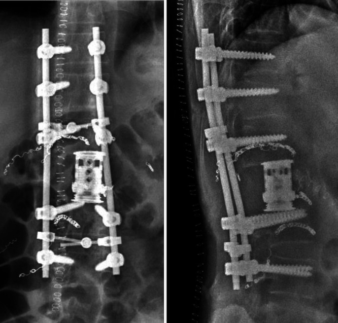
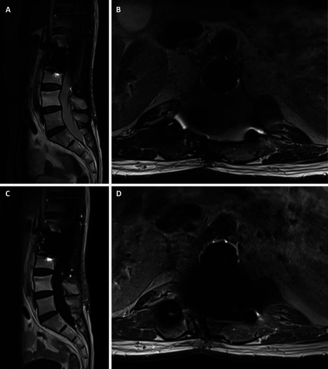
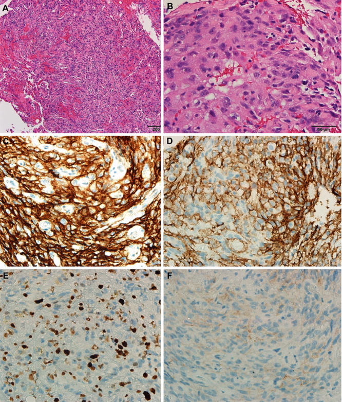
Observations: A 49-year-old man presented with lower back pain and numbness in both lower extremities. Lumbar spine magnetic resonance imaging revealed an L2 pathological fracture with epidural and paraspinal invasion. The patient had undergone a first palliative decompression and fixation surgery, and the diagnosis turned out to be a World Health Organization grade III anaplastic meningioma based on histopathology. The tumor had progressed after first operation and radiation therapy, and the patient was referred to the authors' institute for excision. The patient had an uneventful postoperative course after a revisional total en bloc spondylectomy of L2.
Lessons: The authors present a rare case of PIM of the vertebrae with epidural and paraspinal invasion. Careful preoperative assessment and surgical planning is crucial for successful patient management.
Keywords: CT = computed tomography; MRI = magnetic resonance imaging; PEM = primary extradural meningioma; PIM = primary intraosseous meningioma; WHO = World Health Organization; anaplastic meningioma; primary extradural meningioma; primary intraosseous meningioma; primary spine tumor; total en bloc spondylectomy.
© 2021 The authors.
Conflict of interest statement
Disclosures The authors report no conflict of interest concerning the materials or methods used in this study or the findings specified in this paper.
Figures


Similar articles
-
Metastatic meningioma to the eleventh dorsal vertebral body: total en bloc spondylectomy. Case report and review of the literature.Neurocirugia (Astur). 2006 Jun;17(3):240-9. doi: 10.1016/s1130-1473(06)70346-3. Neurocirugia (Astur). 2006. PMID: 16855782
-
Asymptomatic Intraosseous Meningioma of the Humerus: A Case Report and Review of the Literature.Cureus. 2022 Dec 16;14(12):e32590. doi: 10.7759/cureus.32590. eCollection 2022 Dec. Cureus. 2022. PMID: 36654535 Free PMC article.
-
A Primary Intraosseous Meningioma: A Rare Case of Malignancy with High Proliferative Ability.J Neurol Surg Rep. 2023 Sep 27;84(3):e103-e108. doi: 10.1055/a-2161-7710. eCollection 2023 Jul. J Neurol Surg Rep. 2023. PMID: 37901278 Free PMC article.
-
Atypical Intracranial Meningioma with Metastasis to C7 Vertebral Body: A Case Report.World Neurosurg. 2019 Feb;122:593-598. doi: 10.1016/j.wneu.2018.11.067. Epub 2018 Nov 20. World Neurosurg. 2019. PMID: 30465962 Review.
-
Primary extradural meningioma presenting as a neck mass: Case report and review of the literature.Head Neck. 2015 Aug;37(8):E92-5. doi: 10.1002/hed.23874. Epub 2015 Jun 16. Head Neck. 2015. PMID: 25251307 Review.
Cited by
-
Spinal meningiomas.Neurooncol Adv. 2023 Jun 3;5(Suppl 1):i112-i121. doi: 10.1093/noajnl/vdad013. eCollection 2023 May. Neurooncol Adv. 2023. PMID: 37287574 Free PMC article.
References
-
- Ward AL, Risman A, Segar S, Sharma S, Vender JR. Atypical intracranial meningioma with metastasis to C7 vertebral body: a case report. World Neurosurg. 2019;122:593–598. - PubMed
-
- Lang FF, Macdonald OK, Fuller GN, DeMonte F. Primary extradural meningiomas: a report on nine cases and review of the literature from the era of computerized tomography scanning. J Neurosurg. 2000;93(6):940–950. - PubMed
-
- Elder JB, Atkinson R, Zee CS, Chen TC. Primary intraosseous meningioma. Neurosurg Focus. 2007;23(4):E13. - PubMed
Publication types
LinkOut - more resources
Full Text Sources
Research Materials
