Review
. 2022 Jul 11;13(4):1196-1214.
doi: 10.14336/AD.2022.0109.

Targeting the Hepatic Microenvironment to Improve Ischemia/Reperfusion Injury: New Insights into the Immune and Metabolic Compartments

Affiliations
Review

Targeting the Hepatic Microenvironment to Improve Ischemia/Reperfusion Injury: New Insights into the Immune and Metabolic Compartments

Fengqiang Gao et al. Aging Dis. .

Abstract

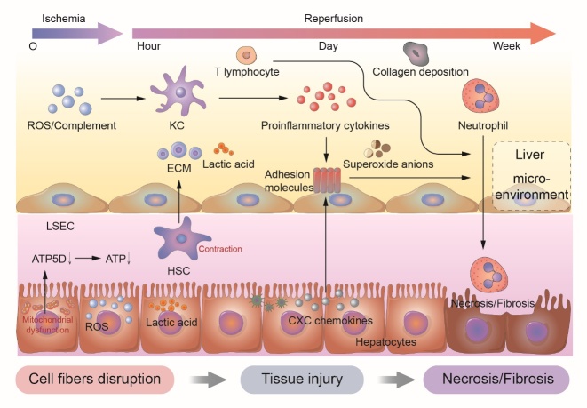
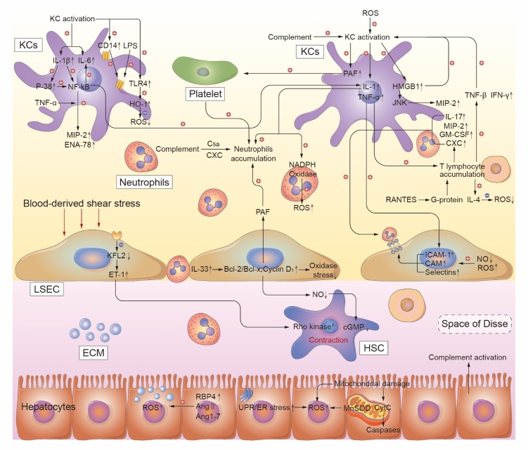
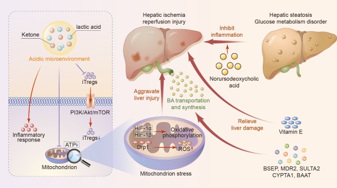
Hepatic ischemia/reperfusion injury (IRI) is mainly characterized by high activation of immune inflammatory responses and metabolic responses. Understanding the molecular and metabolic mechanisms underlying development of hepatic IRI is critical for developing effective therapies for hepatic IRI. Recent advances in research have improved our understanding of the pathogenesis of IRI. During IRI, hepatocyte injury and inflammatory responses are mediated by crosstalk between the immune cells and metabolic components. This crosstalk can be targeted to treat or reverse hepatic IRI. Thus, a deep understanding of hepatic microenvironment, especially the immune and metabolic responses, can reveal new therapeutic opportunities for hepatic IRI. In this review, we describe important cells in the liver microenvironment (especially non-parenchymal cells) that regulate immune inflammatory responses. The role of metabolic components in the diagnosis and prevention of hepatic IRI are discussed. Furthermore, recent updated therapeutic strategies based on the hepatic microenvironment, including immune cells and metabolic components, are highlighted.

Keywords: hepatic microenvironment; immune cell; inflammatory response; ischemia/reperfusion injury; metabolic compartment; therapeutic strategies.


Conflict of interest statement

Conflicts of Interest The authors declare no competing interests.

Figures

Figure 1.
Figure 1.
The regulation of liver microenvironment during different periods of hepatic ischemia/reperfusion injury. A summary of the immune components of liver microenvironment during the continuous phase of ischemia/reperfusion injury is manifested. LSEC, liver sinusoidal endothelial cell; HSC, hepatic stellate cell; KC, kupffer cell; ROS, reactive oxygen species; ECM, extracellular matrix.
Figure 2.
Figure 2.
The regulation of liver microenvironment components, including hepatic parenchymal cells, hepatic non-parenchymal cells (hepatic stellate cells, Kupffer cells, sinusoidal endothelial cells, neutrophils and lymphocytes), and extracellular matrix, during hepatic ischemia/reperfusion injury. A summary of the specific molecular mechanisms regulating hepatocytes and interactions in the liver microenvironment are shown. LSEC, liver sinusoidal endothelial cell; HSC, hepatic stellate cell; KC, Kupffer cell; ROS, reactive oxygen species; ECM, extracellular matrix; IL-1, interleukin 1; IL-6, interleukin 6; IL-17, interleukin 17; IL-33, interleukin 33; IL-1β, interleukin 1β; HMGB1, high-mobility group box 1; IAC, inflammation associated cytokine; TNF-α, tumor necrosis factor α; PAF, platelet activating factor; MIP-2, macrophage inflammatory protein 2; ENA-78, epithelial neutrophil activating protein 78; NF-κB, nuclear factor κB; TLR4, Toll like receptor 4; LPS, lipopolysaccharide; HO-1, heme oxygenase-1; RANTES, regulated upon activation normal T cell expressed and secreted factor; VEGF, vascular endothelial growth factor; IFN-γ, interferon γ; GM-CSF, granulocyte-macrophage colony-stimulating factor; ICAM-1, intercellular adhesion molecule-1; Bcl-2/Bcl-x, B cell lymphoma 2/x; TXA2, thromboxane; KFL2, kruppel like transcription factor 2; ET-1, endothelin 1; JNK, N terminal kinase; CAM, cell adhesion molecule; cGMP, cyclic guanosine monophosphate; RBP4, retinol binding protein; Ang 1-7, angiotensin 1-7; Ang Ⅱ, angiotensin Ⅱ; MnSOD, manganese containing superoxide dismutase; CytC, cytochrome C.
Figure 3.
Figure 3.
Metabolic compartments regulate the process of hepatic ischemia/reperfusion injury. A summary of metabolic components of the liver microenvironment during phase of ischemia/reperfusion injury is manifested. iTregs, inducible regulatory T cells; ATP, adenosine triphosphate; BA, bile acid; Drp1, dynamin-related protein 1; HIF-1α, hypoxia-inducible factor-1.

References

    1. de Groot H, Rauen U (2007). Ischemia-reperfusion injury: processes in pathogenetic networks: a review. Transplant Proc, 39:481-484. - PubMed
    1. Binder A, Ali A, Chawla R, Aziz HA, Abbate A, Jovin IS (2015). Myocardial protection from ischemia-reperfusion injury post coronary revascularization. Expert Rev Cardiovasc Ther, 13:1045-1057. - PubMed
    1. Ginsberg MD (2016). Expanding the concept of neuroprotection for acute ischemic stroke: The pivotal roles of reperfusion and the collateral circulation. Prog Neurobiol, 145-146:46-77. - PubMed
    1. Gonzalez LM, Moeser AJ, Blikslager AT (2015). Animal models of ischemia-reperfusion-induced intestinal injury: progress and promise for translational research. Am J Physiol Gastrointest Liver Physiol, 308:G63-75. - PMC - PubMed
    1. Zhai Y, Busuttil RW, Kupiec-Weglinski JW (2011). Liver ischemia and reperfusion injury: new insights into mechanisms of innate-adaptive immune-mediated tissue inflammation. Am J Transplant, 11:1563-1569. - PMC - PubMed

LinkOut - more resources