Intracranial tuberculoma: a rare complication of extrapulmonary tuberculosis. Illustrative case
- PMID: 35855351
- PMCID: PMC9257396
- DOI: 10.3171/CASE2291
Intracranial tuberculoma: a rare complication of extrapulmonary tuberculosis. Illustrative case
Abstract
Background: Intracranial tuberculomas are rare entities commonly seen only in low- to middle-income countries where tuberculosis remains endemic. Furthermore, following adequate treatment, the development of intracranial spread is uncommon in the absence of immunosuppression.
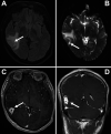
Observations: A 22-year-old man with no history of immunosuppression presented with new-onset seizures in the setting of miliary tuberculosis status post 9 months of antitubercular therapy. Following a 2-month period of remission, he presented with new-onset tonic-clonic seizures. Magnetic resonance imaging demonstrated interval development of a mass concerning for an intracranial tuberculoma. After resection, pathological analysis of the mass revealed caseating granulomas within the multinodular lesion, consistent with intracranial tuberculoma. The patient was discharged after the reinitiation of antitubercular medications along with a steroid taper.
Lessons: To the best of the authors' knowledge, this case represents the first instance of intracranial tuberculoma occurring after the initial resolution of a systemic tuberculosis infection. The importance of retaining a high level of suspicion when evaluating these patients for seizure etiology is crucial because symptoms are rapidly responsive to resection of intracranial tuberculoma masses. Furthermore, it is imperative for surgeons to recognize the isolation steps necessary when managing these patients within the operating theater and inpatient settings.
Keywords: CNS = central nervous system; CSF = cerebrospinal fluid; CT = computed tomography; DWI = diffusion-weighted imaging; MRI = magnetic resonance imaging; extrapulmonary tuberculosis; intracranial; tuberculoma.
© 2022 The authors.
Conflict of interest statement
Disclosures The authors report no conflict of interest concerning the materials or methods used in this study or the findings specified in this paper.
Figures


References
-
- Garg RK, Sharma R, Kar AM, et al. Neurological complications of miliary tuberculosis. Clin Neurol Neurosurg. 2010;112(3):188–192. - PubMed
-
- Bagga A, Kalra V, Ghai OP. Intracranial tuberculoma. Evaluation and treatment. Clin Pediatr (Phila) 1988;27(10):487–490. - PubMed
-
- Kioumehr F, Dadsetan MR, Rooholamini SA, Au A. Central nervous system tuberculosis: MRI. Neuroradiology. 1994;36(2):93–96. - PubMed
-
- Rajshekhar V. Surgery for brain tuberculosis: a review. Acta Neurochir (Wien) 2015;157(10):1665–1678. - PubMed
Publication types
LinkOut - more resources
Full Text Sources

