

Successful endovascular treatment of a ruptured bihemispheric posterior inferior cerebellar artery aneurysm: illustrative case
- PMID: 35855413
- PMCID: PMC9265170
- DOI: 10.3171/CASE21367
Successful endovascular treatment of a ruptured bihemispheric posterior inferior cerebellar artery aneurysm: illustrative case
Abstract
Background: Normal posterior inferior cerebellar artery (PICA) anatomy is highly variable, but bihemispheric PICA crossing the midline to supply the vascular territory of bilateral cerebellar hemisphere is rare. Herein, the authors reported a rare case of ruptured aneurysm that was associated with bihemispheric PICA and successfully treated endovascularly.
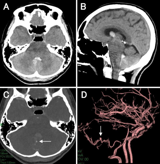
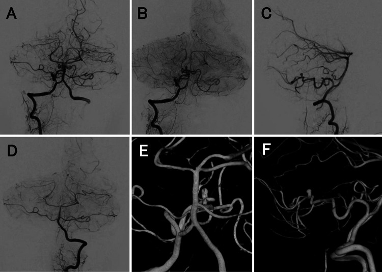
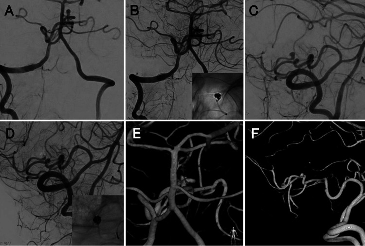
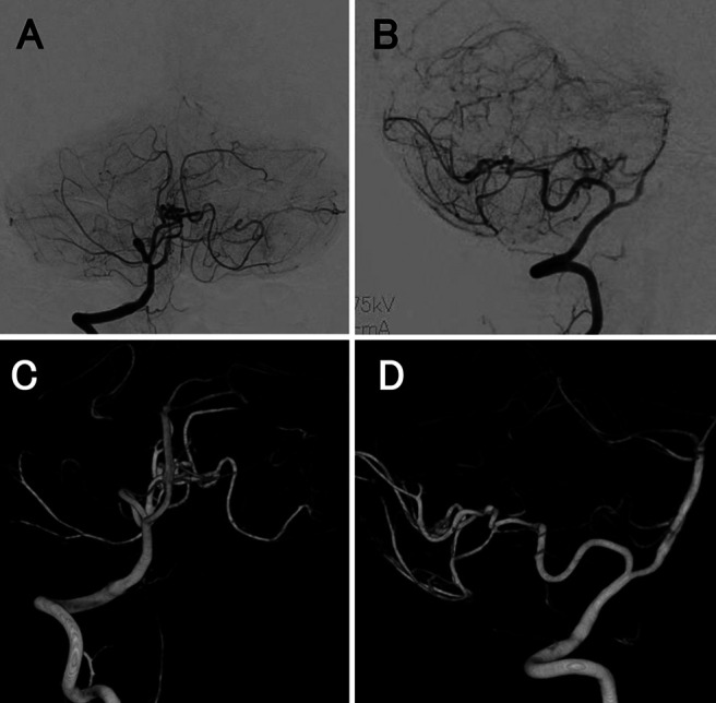
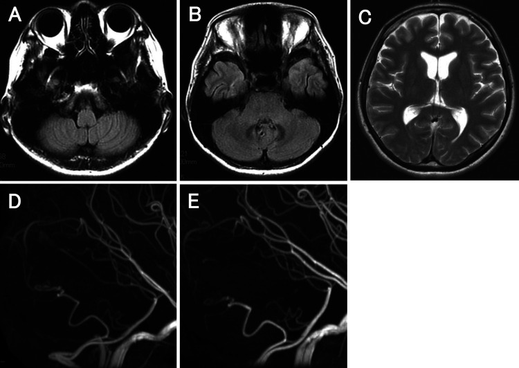
Observations: A 46-year-old woman presented with sudden headache and loss of consciousness because of an intraventricular hemorrhage due to a ruptured aneurysm that was associated with the bihemispheric PICA. Angiography revealed that the aneurysm was located at the bifurcation between the bihemispheric PICA and the bilateral distal PICA. The ruptured aneurysm was successfully occluded using coil embolization, which preserved the parent artery with no procedural-related complication.
Lessons: To the best of the authors' knowledge, this was the first report of a ruptured aneurysm associated with bihemispheric PICA being successfully treated endovascularly. Aneurysm formation may be accelerated by hemodynamic stress and vascular fragility. For neurosurgeons and neurointerventionalists, it is important to understand the anatomical variation of PICA, especially bihemispheric PICA, which is a potential risk factor for a fatal stroke.
Keywords: CT = computed tomography; CTA = CT angiography; DSA = digital subtraction angiography; MRI = magnetic resonance imaging; PICA = posterior inferior cerebellar artery; VA = vertebral artery; bihemispheric PICA; coil embolization; intraventricular hemorrhage; ruptured aneurysm.
© 2021 The authors.
Conflict of interest statement
Disclosures The authors report no conflict of interest concerning the materials or methods used in this study or the findings specified in this paper.
Figures
References
-
- Hudgins RJ, Day AL, Quisling RG, Rhoton AL, Jr, Sypert GW, Garcia-Bengochea F. Aneurysms of the posterior inferior cerebellar artery. A clinical and anatomical analysis. J Neurosurg. 1983;58(3):381–387. - PubMed
-
- Locksley HB. Natural history of subarachnoid hemorrhage, intracranial aneurysms and arteriovenous malformations. Based on 6368 cases in the cooperative study. J Neurosurg. 1966;25(2):219–239. - PubMed
-
- Ishikawa T, Suzuki A, Yasui N. Distal posterior inferior cerebellar aneurysms: report of 12 cases. Neurol Med Chir (Tokyo) 1990;30(2):100–108. - PubMed
-
- Lasjaunias P, Vallee B, Person H, Ter Brugge K, Chiu M. The lateral spinal artery of the upper cervical spinal cord. Anatomy, normal variations, and angiographic aspects. J Neurosurg. 1985;63(2):235–241. - PubMed
Publication types
LinkOut - more resources
Full Text Sources
