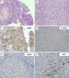
Multifocal primary central nervous system Ewing sarcoma presenting with intracranial hemorrhage and leptomeningeal dissemination: illustrative case
- PMID: 35855436
- PMCID: PMC9241201
- DOI: 10.3171/CASE2042
Multifocal primary central nervous system Ewing sarcoma presenting with intracranial hemorrhage and leptomeningeal dissemination: illustrative case
Abstract
Background: Ewing sarcoma is a neoplasm within the family of small round blue cell tumors and most frequently arises from skeletal bone. Primary involvement of the central nervous system in these lesions is extremely rare, with an incidence of 1%.
Observations: A case is presented of a 34-year-old man who presented with left facial numbness, multiple intracranial lesions, a lumbar intradural lesion, and diffuse spinal leptomeningeal involvement. A lumbar laminectomy and biopsy were performed, which revealed the diagnosis of extraskeletal Ewing sarcoma/primitive neuroectodermal tumor. The patient had a rapidly progressive clinical decline despite total neuroaxis radiation and multiple lines of chemotherapeutic treatments, eventually dying from his disease and its sequelae 6 months after diagnosis.
Lessons: The authors' review of 40 cases in the literature revealed only 2 patients with isolated intraaxial cranial lesions, 4 patients with cranial and spine involvement, and an additional 34 patients with spine lesions. The unique characteristics of this patient's case, including his presentation with diffuse disease and pathology that included a rare V600E BRAF mutation, are discussed in the context of the available literature.
Keywords: BRAF; CNS = central nervous system; CSF = cerebrospinal fluid; CT = computed tomography; ES = Ewing sarcoma; Ewing sarcoma; GFAP = glial fibrillary acidic protein; MRI = magnetic resonance imaging; cPNET = central primitive neuroectodermal tumor; intracranial; oncology; spine.
© 2021 The authors.
Conflict of interest statement
Disclosures Dr. Perkins is employed by Washington University and is a paid member of the medical advisory committee for Mevion Medical Systems, Inc. Dr. Chicoine received grants from IMRIS, Inc., The Head for the Cure Foundation, Mrs. Carol Rossfeld, The Alex & Alice Aboussie Family Charitable Foundation, and Subcortical Surgery Group Research Grant Program, outside of the submitted work.
Figures


Similar articles
-
Primary intracranial dural-based Ewing sarcoma/peripheral primitive neuroectodermal tumor mimicking a meningioma: A rare tumor with review of literature.Asian J Neurosurg. 2017 Jul-Sep;12(3):351-357. doi: 10.4103/1793-5482.185060. Asian J Neurosurg. 2017. PMID: 28761507 Free PMC article. Review.
-
Primary intradural extraosseous Ewing sarcoma of the spine: case report and literature review.Neurosurgery. 2011 Oct;69(4):E995-9. doi: 10.1227/NEU.0b013e318223b7c7. Neurosurgery. 2011. PMID: 21572359 Review.
-
Primary extraskeletal intradural Ewing sarcoma with acute hemorrhage: a case report and review of the literature.J Med Case Rep. 2024 Mar 9;18(1):144. doi: 10.1186/s13256-024-04384-8. J Med Case Rep. 2024. PMID: 38459600 Free PMC article.
-
Intracranial Ewing sarcoma: four pediatric examples.Childs Nerv Syst. 2018 Mar;34(3):441-448. doi: 10.1007/s00381-017-3684-7. Epub 2017 Dec 28. Childs Nerv Syst. 2018. PMID: 29285586 Free PMC article.
-
A case report on non-metastatic Ewing sarcoma of the lumbar spine in a young patient.Cancer Rep (Hoboken). 2022 Nov;5(11):e1725. doi: 10.1002/cnr2.1725. Epub 2022 Oct 3. Cancer Rep (Hoboken). 2022. PMID: 36193025 Free PMC article.
Cited by
-
Primary and metastatic cerebral Ewing's sarcoma: A case report about a rare entity and literature review.Surg Neurol Int. 2024 Oct 11;15:367. doi: 10.25259/SNI_316_2024. eCollection 2024. Surg Neurol Int. 2024. PMID: 39524588 Free PMC article.
-
Case report: Primary intracranial EWs/PNET in adults: Clinical experience and literature review.Front Oncol. 2022 Oct 13;12:1035800. doi: 10.3389/fonc.2022.1035800. eCollection 2022. Front Oncol. 2022. PMID: 36313718 Free PMC article.
-
Intracranial peripheral primitive neuroectodermal tumor presenting as neurosurgical emergency: A report of two cases.J Neurosci Rural Pract. 2023 Jan-Mar;14(1):119-122. doi: 10.25259/JNRP-2022-1-33. Epub 2022 Dec 15. J Neurosci Rural Pract. 2023. PMID: 36891115 Free PMC article.
-
Primary Intracranial Ewing Sarcoma With EWSR1-FLI1 Gene Translocation Mimicking a Meningioma and a Multidisciplinary Therapeutic Approach: A Case Report and Systematic Review of Literatures.Brain Tumor Res Treat. 2023 Oct;11(4):281-288. doi: 10.14791/btrt.2023.0030. Brain Tumor Res Treat. 2023. PMID: 37953453 Free PMC article.
-
The Role of Neuroaxis Irradiation in the Treatment of Intraspinal Ewing Sarcoma: A Review and Meta-Analysis.Cancers (Basel). 2022 Feb 25;14(5):1209. doi: 10.3390/cancers14051209. Cancers (Basel). 2022. PMID: 35267515 Free PMC article. Review.
References
-
- Esiashvili N, Goodman M, Marcus RB., Jr Changes in incidence and survival of Ewing sarcoma patients over the past 3 decades: Surveillance Epidemiology and End Results data. J Pediatr Hematol Oncol. 2008;30(6):425–430. - PubMed
-
- Balamuth NJ, Womer RB. Ewing’s sarcoma. Lancet Oncol. 2010;11(2):184–192. - PubMed
-
- Agrawal A, Dulani R, Mahadevan A, et al. Primary Ewing’s sarcoma of the frontal bone with intracranial extension. J Cancer Res Ther. 2009;5(3):208–209. - PubMed
-
- Antonelli M, Caltabiano R, Chiappetta C, et al. Primary peripheral PNET/Ewing’s sarcoma arising in the meninges, confirmed by the presence of the rare translocation t(21;22) (q22;q12) Neuropathology. 2011;31(5):549–555. - PubMed
-
- Khalatbari MR, Jalaeikhoo H, Moharamzad Y. Primary intradural extraosseous Ewing’s sarcoma of the lumbar spine presenting with acute bleeding. Br J Neurosurg. 2013;27(6):840–841. - PubMed
Publication types
LinkOut - more resources
Full Text Sources
Research Materials
Miscellaneous

