Review
. 2022 Jul;12(7):220054.
doi: 10.1098/rsob.220054. Epub 2022 Jul 20.

Methods to study folding of alpha-helical membrane proteins in lipids

Affiliations
Review

Methods to study folding of alpha-helical membrane proteins in lipids

Nicola J Harris et al. Open Biol. 2022 Jul.

Abstract

How alpha-helical membrane proteins fold correctly in the highly hydrophobic membrane interior is not well understood. Their folding is known to be highly influenced by the lipids within the surrounding bilayer, but the majority of folding studies have focused on detergent-solubilized protein rather than protein in a lipid environment. There are different ways to study folding in lipid bilayers, and each method has its own advantages and disadvantages. This review will discuss folding methods which can be used to study alpha-helical membrane proteins in bicelles, liposomes, nanodiscs or native membranes. These folding methods include in vitro folding methods in liposomes such as denaturant unfolding studies, and single-molecule force spectroscopy studies in bicelles, liposomes and native membranes. This review will also discuss recent advances in co-translational folding studies, which use cell-free expression with liposomes or nanodiscs or are performed in vivo with native membranes.

Keywords: folding; lipids; membrane; protein.


Conflict of interest statement

The authors declare that there are no competing interests associated with this manuscript.

Figures
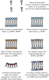
Figure 1.
Figure 1.
Different lipids form bilayers with different properties. Lipids can be bilayer-forming (e.g. DMPC, DOPC, DOPG) or non-bilayer forming (e.g. DOPE). They can have different headgroups which can be charged or neutral, and can have different chains which can be saturated or unsaturated, or branched. Mixing together different types of lipids produces bilayers with different chemical and physical properties.
Figure 2.
Figure 2.
Membrane mimics for membrane protein folding studies. Different membrane mimetics can be used during folding studies. More straightforward systems include detergents or mixed micelles of detergents and lipids. To study folding in lipids, bicelles or membrane scaffold protein (MSP)-based nanodiscs can be used which contain small lipid bilayer sections, with a short-chain detergent around the edges in bicelles, or MSP around the edges in nanodiscs. More advanced folding studies use liposomes, as either small (SUV), large (LUV) or giant (GUV) unilamellar vesicles which contain an inner compartment. This compartmentalization means that liposomes can be used to assess protein topology and for functional assays such as transport assays. The most native lipid system for folding studies is a native lipid extract, or alternatively a novel co-polymer nanodisc can be used which extracts a patch of native lipids from the membrane.
Figure 3.
Figure 3.
Denaturant folding studies in liposomes. Reversible unfolding of membrane proteins in denaturant can give information on unfolding kinetics and thermodynamics [17]. A fully folded protein is reconstituted into liposomes and a denaturant such as urea is added to partially unfold the protein. Removal of the denaturant can allow the protein to refold. Unfolding and refolding are observed using a variety of techniques, such as by measuring the change in secondary structure by circular dichroism, fluorescence (either intrinsic or via a fluorescent label), or by a protease protection assay. Free energies of unfolding (ΔGU) and refolding (ΔGF) can be calculated from the equilibrium constant of the fraction of unfolded/folded proteins.
Figure 4.
Figure 4.
Single-molecule mechanical folding methods. In (a), a typical atomic force microscopy (AFM) set-up is shown. A membrane protein sample either in native membranes or reconstituted into liposomes is deposited onto a piezo stage is approached by a sharp cantilever which scans the surface of the sample. The force generated/experienced deflects the cantilever which in turn is detected by a laser and photodiode detector. When the cantilever is retracted from the surface at a constant velocity, a bound membrane protein unfolds in a stepwise manner which is characteristic of the proteins' intrinsic stability. A ‘saw-tooth’ pattern of unfolding is generated where each peak is fit to the worm-like chain (WLC) model of polymer elasticity. The end-to-end length of the unfolded protein or contour length (LC) is calculated from WLC for each structural segment which unfolds at the given pulling velocity. The LC can be converted into the number of amino acids unfolded at each event. Alternatively (b), a magnetic tweezer set-up can unfold a membrane protein along the plane of the membrane which has been reconstituted into a bicelle or liposome. DNA nano-tethers bind the protein termini to a functionalized magnetic bead and stage. When the pulling velocity is increased, the protein is stretched along the membrane and an unfolding intermediate is observed. This unfolding can be reversible, the protein refolding as the force is decreased.
Figure 5.
Figure 5.
Cell-free expression of membrane proteins. Much of the data on membrane protein co-translational folding come from studies which express the protein cell-free. In vitro transcription/translation (IVTT) machinery is produced recombinantly or extracted from a host cell system and mixed with a membrane mimic and the gene for the membrane protein of choice (DNA or RNA). Optional extras to follow the folding of the protein can be added into the reaction mixture, such as radiolabels, fluorescent unnatural amino acids or chaperones. Correctly folded proteins in the membrane mimic can be isolated from the reaction mixture and aggregate/misfolded protein. A sucrose gradient is often used for this when the protein is expressed into liposomes.
Figure 6.
Figure 6.
Arrest peptide studies for membrane protein folding. (a) The LebB construct used for arrest peptide (AP) force measurements from [125]. A leucine/alanine (L/A) helix was cloned upstream of the polytopic membrane protein to maintain the proteins native N-in orientation. The predicted +ΔGapp was calculated for each helix, and a test TM helix was selected from the proteins CaiT, NhaA, EmrD, BtuC and GlpT. Downstream of this at variable length (L) is a SecM AP peptide. (b) Schematic of the experimental set-up. The ribosome translates the LepB construct and the helices insert into the membrane via the translocon. Once the AP stalls, the force produced by the partitioning of the test helix into the membrane can release the AP. An fFL value is calculated by the fraction of full-length versus truncated protein produced as determined by western blot.

References

    1. Kaushal S, Khorana HG. 1994. Structure and function in rhodopsin. 7. Point mutations associated with autosomal dominant retinitis pigmentosa. Biochemistry 33, 6121-6128. (10.1021/bi00186a011) - DOI - PubMed
    1. Bose SJ, et al. 2020. Towards next generation therapies for cystic fibrosis: folding, function and pharmacology of CFTR. J. Cyst. Fibros. 19(Suppl 1), S25-S32. (10.1016/j.jcf.2019.12.009) - DOI - PMC - PubMed
    1. Shishido H, Yoon JS, Yang Z, Skach WR. 2020. CFTR trafficking mutations disrupt cotranslational protein folding by targeting biosynthetic intermediates. Nat. Commun. 11, 4258. (10.1038/s41467-020-18101-8) - DOI - PMC - PubMed
    1. Kuhn A, Kiefer D. 2017. Membrane protein insertase YidC in bacteria and archaea. Mol. Microbiol. 103, 590-594. (10.1111/mmi.13586) - DOI - PubMed
    1. Cymer F, von Heijne G, White SH. 2015. Mechanisms of integral membrane protein insertion and folding. J. Mol. Biol. 427, 999-1022. (10.1016/j.jmb.2014.09.014) - DOI - PMC - PubMed

Publication types

LinkOut - more resources