. 2022 Sep;48(3):154.
doi: 10.3892/or.2022.8366. Epub 2022 Jul 20.

Defucosylated mouse‑dog chimeric anti‑HER2 monoclonal antibody exerts antitumor activities in mouse xenograft models of canine tumors

Affiliations

Defucosylated mouse‑dog chimeric anti‑HER2 monoclonal antibody exerts antitumor activities in mouse xenograft models of canine tumors

Hiroyuki Suzuki et al. Oncol Rep. 2022 Sep.

Abstract

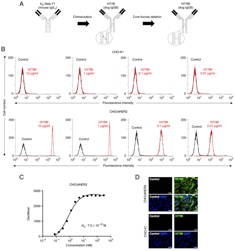
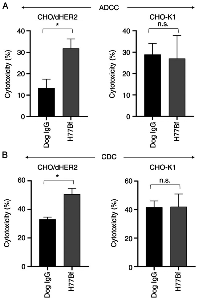
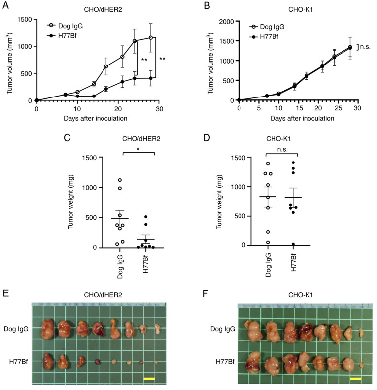
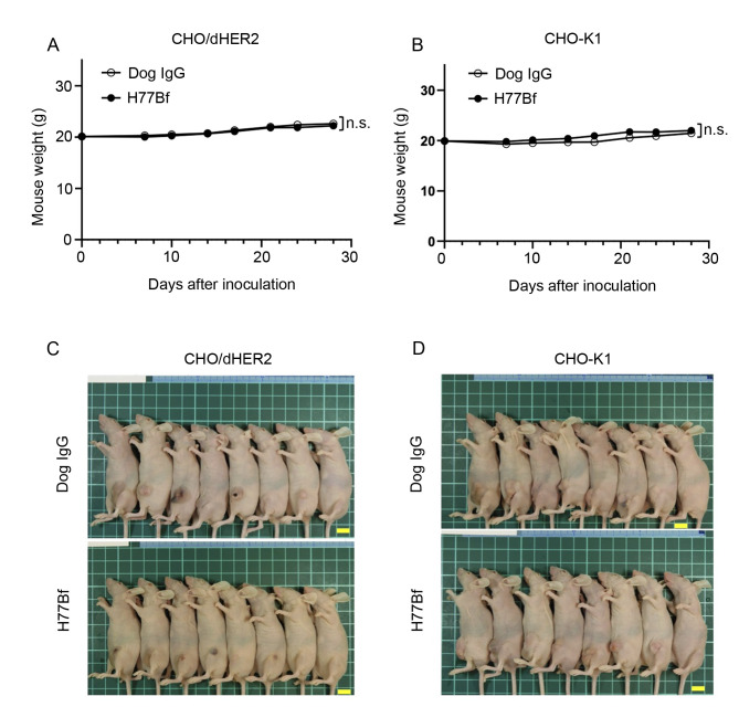
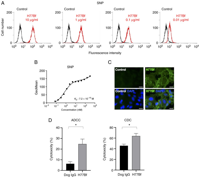
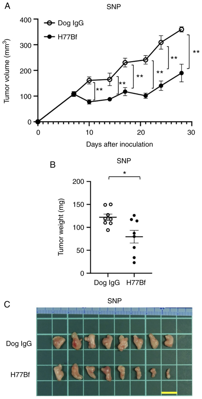
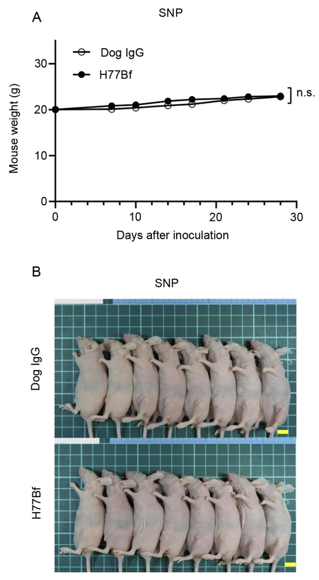
Human epidermal growth factor receptor 2 (HER2) overexpression has been reported in various types of cancer, including breast, gastric, lung, colorectal and pancreatic cancer. A humanized anti‑HER2 monoclonal antibody (mAb), trastuzumab, has been shown to improve survival of patients in HER2‑positive breast and gastric cancer. An anti‑HER2 mAb, H2Mab‑77 (mouse IgG1, kappa) was previously developed. In the present study, a defucosylated version of mouse‑dog chimeric anti‑HER2 mAb (H77Bf) was generated. H77Bf possesses a high binding‑affinity [a dissociation constant (KD): 7.5x10‑10 M, as determined by flow cytometric analysis] for dog HER2‑overexpressed CHO‑K1 (CHO/dHER2) cells. H77Bf highly exerted antibody‑dependent cellular cytotoxicity (ADCC) and complement‑dependent cytotoxicity (CDC) for CHO/dHER2 cells by canine mononuclear cells and complement, respectively. Moreover, administration of H77Bf significantly suppressed the development of CHO/dHER2 xenograft tumor in mice compared with the control dog IgG. H77Bf also possesses a high binding‑affinity (KD: 7.2x10‑10 M) for a canine mammary gland tumor cell line (SNP), and showed high ADCC and CDC activities for SNP cells. Intraperitoneal administration of H77Bf in mouse xenograft models of SNP significantly suppressed the development of SNP xenograft tumors compared with the control dog IgG. These results indicated that H77Bf exerts antitumor activities against dHER2‑positive canine cancers, and could be valuable treatment regimen for canine cancers.

Keywords: ADCC; CDC; HER2; antitumor activity; monoclonal antibody.


Conflict of interest statement

The authors declare that they have no competing interests.

Figures

Figure 1.
Figure 1.
Flow cytometry using H77Bf. (A) Production of H77Bf (core-fucose-deficient dog IgGB) from H2Mab-77 (mouse IgG1). (B) CHO-K1 and CHO/dHER2 cells were treated with H77Bf or buffer control, followed by FITC-conjugated anti-dog IgG. (C) Determination of the binding affinity of H77Bf using flow cytometry for CHO/dHER2 cells. CHO/dHER2 cells were suspended in serially diluted H77Bf, followed by the addition of FITC-conjugated anti-dog IgG. Fluorescence data were analyzed using the EC800 Cell Analyzer. (D) Immunocytochemistry using H77Bf. CHO-K1 and CHO/dHER2 cells were incubated with buffer control or 10 µg/ml H77Bf for 1 h, followed by the incubation with Alexa Fluor 488-conjugated anti-dog IgG and DAPI for 45 min. Fluorescent images were acquired using a fluorescent microscope BZ-X800. Scale bars, 20 µm.
Figure 2.
Figure 2.
Evaluation of ADCC and CDC elicited by H77Bf. (A) ADCC elicited by H77Bf and control dog IgG targeting CHO/dHER2 and CHO-K1 cells. (B) CDC elicited by H77Bf and control dog IgG targeting CHO/dHER2 and CHO-K1 cells Values are presented as the mean ± SEM. (*P<0.05; Welch's t- test). ADCC, antibody-dependent cellular cytotoxicity; CDC, complement-dependent cytotoxicity; n.s., not significant.
Figure 3.
Figure 3.
Antitumor activity of H77Bf. (A and B) Evaluation of tumor volume in (A) CHO/dHER2 and (B) CHO-K1 ×enograft models. CHO/dHER2 and CHO-K1 cells (5×10⁶ cells) were subcutaneously injected into mice. On day 8, 100 µg of H77Bf or control dog IgG were injected intraperitoneally into mice. Additional antibodies were injected on days 14 and 21. Mononuclear cells were also injected surrounding the tumors on days 8, 14 and 21. The tumor volume was measured on days 7, 10, 14, 17, 21, 24 and 28 after the injection. Values are presented as the mean ± SEM. **P<0.01 (ANOVA and Sidak's multiple comparisons test). (C and D) Tumor weight (day 28) was measured from excised xenografts of (C) CHO/dHER2 and (D) CHO-K1. Values are presented as the mean ± SEM. *P<0.05 (Welch's t-test). (E and F) Appearance of resected tumors of (E) CHO/dHER2 and (F) CHO-K1 ×enografts from the control dog IgG and H77Bf treated groups on day 28 (scale bar, 1 cm). n.s., not significant.
Figure 4.
Figure 4.
Body weights and appearance of the mice. (A and B) Body weights of mice implanted with (A) CHO/dHER2 and (B) CHO-K1 ×enografts on days 7, 10, 14, 17, 21, 24 and 28 (ANOVA and Sidak's multiple comparisons test). (C and D) Body appearance of (C) CHO/dHER2 and (D) CHO-K1-implanted mice on day 28 (scale bar, 1 cm). n.s., not significant.
Figure 5.
Figure 5.
Flow cytometry, ADCC and CDC activity of H77Bf against canine mammary gland tumor cell line, SNP cells. (A) SNP cells were treated with H77Bf or buffer control, followed by FITC-conjugated anti-dog IgG. (B) Determination of the binding affinity of H77Bf for SNP cells using flow cytometry. SNP cells were suspended in 100 µl of serially diluted H77Bf, followed by the addition of FITC-conjugated anti-dog IgG. Fluorescence data were collected using the EC800 Cell Analyzer. (C) Immunocytochemistry using H77Bf. SNP cells were incubated with buffer control or 10 µg/ml H77Bf for 1 h, followed by the incubation with Alexa Fluor 488-conjugated anti-dog IgG and DAPI for 45 min. Fluorescent images were acquired using a fluorescent microscope BZ-X800 (scale bars, 20 µm). (D) ADCC and CDC elicited by H77Bf and control dog IgG targeting SNP cells. Values are presented as the mean ± SEM. *P<0.05; Welch's t-test). ADCC, antibody-dependent cellular cytotoxicity; CDC, complement-dependent cytotoxicity.
Figure 6.
Figure 6.
Antitumor activity of H77Bf against SNP xenograft. (A) Evaluation of tumor volume in SNP xenograft models. SNP cells (5×10⁶ cells) were injected subcutaneously into mice. On day 8, 100 µg of H77Bf or control dog IgG in 100 µl PBS were injected intraperitoneally into mice. Additional antibodies were injected on days 14 and 21. Mononuclear cells were also injected surrounding the tumors on days 8, 14 and 21. The tumor volume was measured on days 7, 10, 14, 17, 21, 24 and 28 after the inoculation. Values are presented as the mean ± SEM. **P<0.01 (ANOVA and Sidak's multiple comparisons test). (B) Tumor weight (day 28) was measured from excised SNP xenografts. Values are presented as the mean ± SEM. *P<0.05 (Welch's t-test). (C) Appearance of resected SNP xenografts from the control dog IgG and H77Bf treated groups on day 28 (scale bar, 1 cm).
Figure 7.
Figure 7.
Body weights and appearance of the mice. (A) Body weights of mice implanted with SNP xenografts on days 7, 10, 14, 17, 21, 24 and 28 (ANOVA and Sidak's multiple comparisons test). (B) Body appearance of SNP-implanted mice on day 28 (scale bar, 1 cm). n.s., not significant.

References

    1. Slamon DJ, Clark GM, Wong SG, Levin WJ, Ullrich A, McGuire WL. Human breast cancer: Correlation of relapse and survival with amplification of the HER-2/neu oncogene. Science. 1987;235:177–182. doi: 10.1126/science.3798106. - DOI - PubMed
    1. Slamon DJ, Leyland-Jones B, Shak S, Fuchs H, Paton V, Bajamonde A, Fleming T, Eiermann W, Wolter J, Pegram M, et al. Use of chemotherapy plus a monoclonal antibody against HER2 for metastatic breast cancer that overexpresses HER2. N Engl J Med. 2001;344:783–792. doi: 10.1056/NEJM200103153441101. - DOI - PubMed
    1. Bang YJ, Van Cutsem E, Feyereislova A, Chung HC, Shen L, Sawaki A, Lordick F, Ohtsu A, Omuro Y, Satoh T, et al. Trastuzumab in combination with chemotherapy versus chemotherapy alone for treatment of HER2-positive advanced gastric or gastro-oesophageal junction cancer (ToGA): A phase 3, open-label, randomised controlled trial. Lancet. 2010;376:687–697. doi: 10.1016/S0140-6736(10)61121-X. - DOI - PubMed
    1. Moasser MM, Krop IE. The evolving landscape of HER2 targeting in breast cancer. JAMA Oncol. 2015;1:1154–1161. doi: 10.1001/jamaoncol.2015.2286. - DOI - PubMed
    1. Moasser MM. Two dimensions in targeting HER2. J Clin Oncol. 2014;32:2074–2077. doi: 10.1200/JCO.2014.55.7652. - DOI - PMC - PubMed