. 2022 Aug 30;13(4):e0118322.
doi: 10.1128/mbio.01183-22. Epub 2022 Jul 20.

The Gut Bacterial Community Potentiates Clostridioides difficile Infection Severity

Affiliations

The Gut Bacterial Community Potentiates Clostridioides difficile Infection Severity

Nicholas A Lesniak et al. mBio. .

Abstract

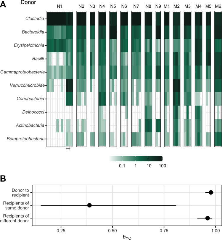
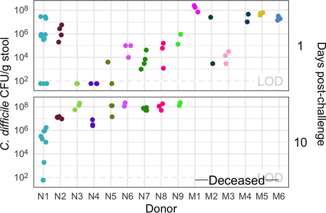
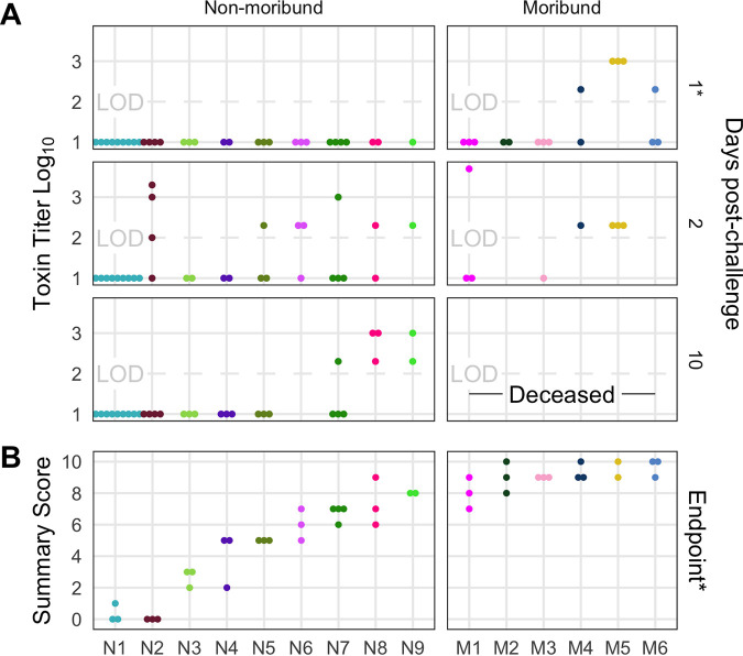
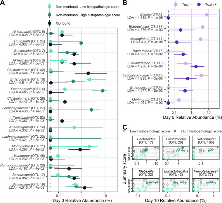
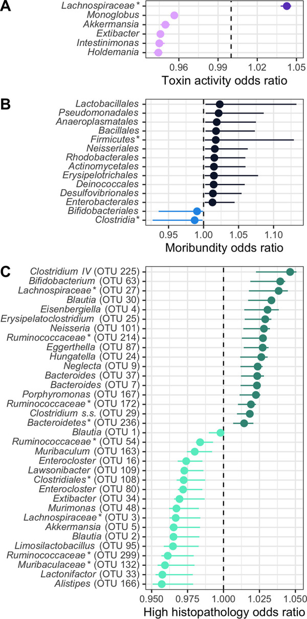
The severity of Clostridioides difficile infections (CDI) has increased over the last few decades. Patient age, white blood cell count, and creatinine levels as well as C. difficile ribotype and toxin genes have been associated with disease severity. However, it is unclear whether specific members of the gut microbiota are associated with variations in disease severity. The gut microbiota is known to interact with C. difficile during infection. Perturbations to the gut microbiota are necessary for C. difficile to colonize the gut. The gut microbiota can inhibit C. difficile colonization through bile acid metabolism, nutrient consumption, and bacteriocin production. Here, we sought to demonstrate that members of the gut bacterial communities can also contribute to disease severity. We derived diverse gut communities by colonizing germfree mice with different human fecal communities. The mice were then infected with a single C. difficile ribotype 027 clinical isolate, which resulted in moribundity and histopathologic differences. The variation in severity was associated with the human fecal community that the mice received. Generally, bacterial populations with pathogenic potential, such as Enterococcus, Helicobacter, and Klebsiella, were associated with more-severe outcomes. Bacterial groups associated with fiber degradation and bile acid metabolism, such as Anaerotignum, Blautia, Lactonifactor, and Monoglobus, were associated with less-severe outcomes. These data indicate that, in addition to the host and C. difficile subtype, populations of gut bacteria can influence CDI disease severity. IMPORTANCE Clostridioides difficile colonization can be asymptomatic or develop into an infection ranging in severity from mild diarrhea to toxic megacolon, sepsis, and death. Models that predict severity and guide treatment decisions are based on clinical factors and C. difficile characteristics. Although the gut microbiome plays a role in protecting against CDI, its effect on CDI disease severity is unclear and has not been incorporated into disease severity models. We demonstrated that variation in the microbiome of mice colonized with human feces yielded a range of disease outcomes. These results revealed groups of bacteria associated with both severe and mild C. difficile infection outcomes. Gut bacterial community data from patients with CDI could improve our ability to identify patients at risk of developing more severe disease and improve interventions that target C. difficile and the gut bacteria to reduce host damage.

Keywords: CDI; Clostridium difficile; human microbiome; humanized mice; microbial ecology.


Conflict of interest statement

The authors declare no conflict of interest.

Figures

FIG 1
FIG 1
Human fecal microbial communities established diverse gut bacterial communities in germfree mice. (A) Relative abundances of the 10 most abundant bacterial classes observed in the feces of previously germfree C57BL/6 mice 14 days postcolonization with human fecal samples (i.e., day 0 relative to C. difficile challenge). Each column of abundances represents an individual mouse. Mice that received the same donor feces are grouped together and labeled above with a letter (“N” for nonmoribund mice and “M” for moribund mice) and number (ordered by mean histopathologic score of the donor group). “+” indicates the mice that did not have detectable C. difficile CFU (Fig. 2). (B) Medians (points) and interquartile ranges (lines) of β diversity (θYC) between an individual mouse and either all others that were inoculated with feces from the same donor or inoculated with feces from a different donor. The β diversity among the same-donor comparison group was significantly less than the β diversity of either the different-donor group or the overall donor community (P < 0.05, calculated by Wilcoxon rank sum test).
FIG 2
FIG 2
All donor groups resulted in C. difficile infection, but with different outcomes. The number of C. difficile CFU per gram of stool was measured the day after challenge with 103 C. difficile RT027 clinical isolate 431 spores and at the end of the experiment, 10 days postchallenge. Each point represents an individual mouse. Mice are grouped by donor and labeled by the donor letter (“N” for nonmoribund mice and “M” for moribund mice) and number (ordered by mean histopathologic score of the donor group). Points are colored by donor group. Mice from donor groups N1 through N6 succumbed to the infection prior to day 10 and were not plated on day 10 postchallenge. LOD, limit of detection. “—Deceased—” indicates mice were deceased at that time point, so no sample was available.
FIG 3
FIG 3
Histopathologic scores and toxin activities varied across donor groups. (A) Fecal toxin activity was detected in some mice post-C. difficile challenge in both moribund and nonmoribund mice. (B) Cecum scored for histopathologic damage from mice at the end of the experiment. Samples were collected for histopathologic scoring on day 10 postchallenge for nonmoribund mice or the day the mouse succumbed to the infection for the moribund group (day 2 or 3 postchallenge). Each point represents an individual mouse. Mice are grouped by donor and labeled by the donor letter (“N” for nonmoribund mice and “M” for moribund mice) and number (ordered by mean histopathologic score of the donor group). Points are colored by donor group. Mice in group N1 that have a summary score of 0 are the mice that did not have detectable C. difficile CFU (Fig. 2). Missing points are from mice that had insufficient fecal sample collected for assaying toxin or cecum for histopathologic scoring. *, significant difference between nonmoribund and moribund groups of mice by Wilcoxon test (P < 0.002).
FIG 4
FIG 4
Individual fecal bacterial community members of the murine gut associated with C. difficile infection outcomes. (A and B) Relative abundance of OTUs at the time of C. difficile challenge (day 0) that varied significantly by the moribundity and histopathologic summary score or with detected toxin by LEfSe analysis. The median (points) and interquartile range (lines) are plotted. (A) Day 0 relative abundances were compared across infection outcome of moribund (colored black) or nonmoribund with either a high histopathologic score (score greater than the median score of 5, colored green) or a low histopathologic summary score (score less than the median score of 5, colored light green). (B) Day 0 relative abundances were compared between mice in which toxin activity was detected (Toxin +, colored dark purple) and which no toxin activity was detected (Toxin −, colored light purple). (C) Day 10 bacterial OTU relative abundances correlated with histopathologic summary score. Data for each mouse are plotted and colored according to their categorization in panel A. Points at the median score of 5 (gray points) were not included in panel A. Spearman’s correlations were statistically significant after Benjamini-Hochberg correction for multiple comparisons. All bacterial groups are ordered by the LDA score. *, the bacterial group was unclassified at lower taxonomic classification ranks.
FIG 5
FIG 5
Fecal bacterial community members of the murine gut at the time of C. difficile infection predicted outcomes of the infection. On the day of infection (day 0), bacterial community members grouped by different classification rank were modeled with logistic regression to predict the infection outcome. The models used the highest taxonomic classification rank without a decrease in performance. Models used all community members, but plotted are those members with a mean odds ratio not equal to 1. The medians (solid points) and interquartile ranges (lines) of the odds ratio are plotted. Bacterial groups are ordered by their odds ratio. *, bacterial group was unclassified at lower taxonomic classification ranks. (A) Bacterial members grouped by genus predicted which mice would have toxin activity detected at any point throughout the infection. Data with a decreased probability of toxin activity are colored light purple, and those with an increased probability of toxin activity are colored dark purple. (B) Bacterial members grouped by order predicted which mice would become moribund. Data with a decreased probability of moribundity are colored light blue, and those with an increased probability of moribundity are colored dark blue. (C) Bacterial members grouped by OTU predicted if the mice would have a high (greater than the median score of 5) or low (less than the median score of 5) histopathologic summary score. Data with a decreased probability of high histopathologic score are colored light green, and those with an increased probability of high histopathologic score are colored dark green.

References

    1. Kelly CP, LaMont JT. 2008. Clostridium difficile—more difficult than ever. N Engl J Med 359:1932–1940. doi:10.1056/NEJMra0707500. - DOI - PubMed
    1. McDonald LC, Gerding DN, Johnson S, Bakken JS, Carroll KC, Coffin SE, Dubberke ER, Garey KW, Gould CV, Kelly C, Loo V, Sammons JS, Sandora TJ, Wilcox MH. 2018. Clinical practice guidelines for Clostridium difficile infection in adults and children: 2017 update by the Infectious Diseases Society of America (IDSA) and Society for Healthcare Epidemiology of America (SHEA). Clin Infect Dis 66:e1–e48. doi:10.1093/cid/cix1085. - DOI - PMC - PubMed
    1. Perry DA, Shirley D, Micic D, Patel CP, Putler R, Menon A, Young VB, Rao K. 2021. External validation and comparison of Clostridioides difficile severity scoring systems. Clin Infect Dis 74:2028–2035. doi:10.1093/cid/ciab737. - DOI - PMC - PubMed
    1. Buffie CG, Bucci V, Stein RR, McKenney PT, Ling L, Gobourne A, No D, Liu H, Kinnebrew M, Viale A, Littmann E, van den Brink MRM, Jenq RR, Taur Y, Sander C, Cross JR, Toussaint NC, Xavier JB, Pamer EG. 2015. Precision microbiome reconstitution restores bile acid mediated resistance to Clostridium difficile. Nature 517:205–208. doi:10.1038/nature13828. - DOI - PMC - PubMed
    1. Britton RA, Young VB. 2014. Role of the intestinal microbiota in resistance to colonization by Clostridium difficile. Gastroenterology 146:1547–1553. doi:10.1053/j.gastro.2014.01.059. - DOI - PMC - PubMed

Publication types

Substances

LinkOut - more resources