. 2022 Aug 31;10(4):e0208322.
doi: 10.1128/spectrum.02083-22. Epub 2022 Jul 20.

The In Vitro Replication Cycle of Achromobacter xylosoxidans and Identification of Virulence Genes Associated with Cytotoxicity in Macrophages

Affiliations

The In Vitro Replication Cycle of Achromobacter xylosoxidans and Identification of Virulence Genes Associated with Cytotoxicity in Macrophages

Adam M Pickrum et al. Microbiol Spectr. .

Abstract

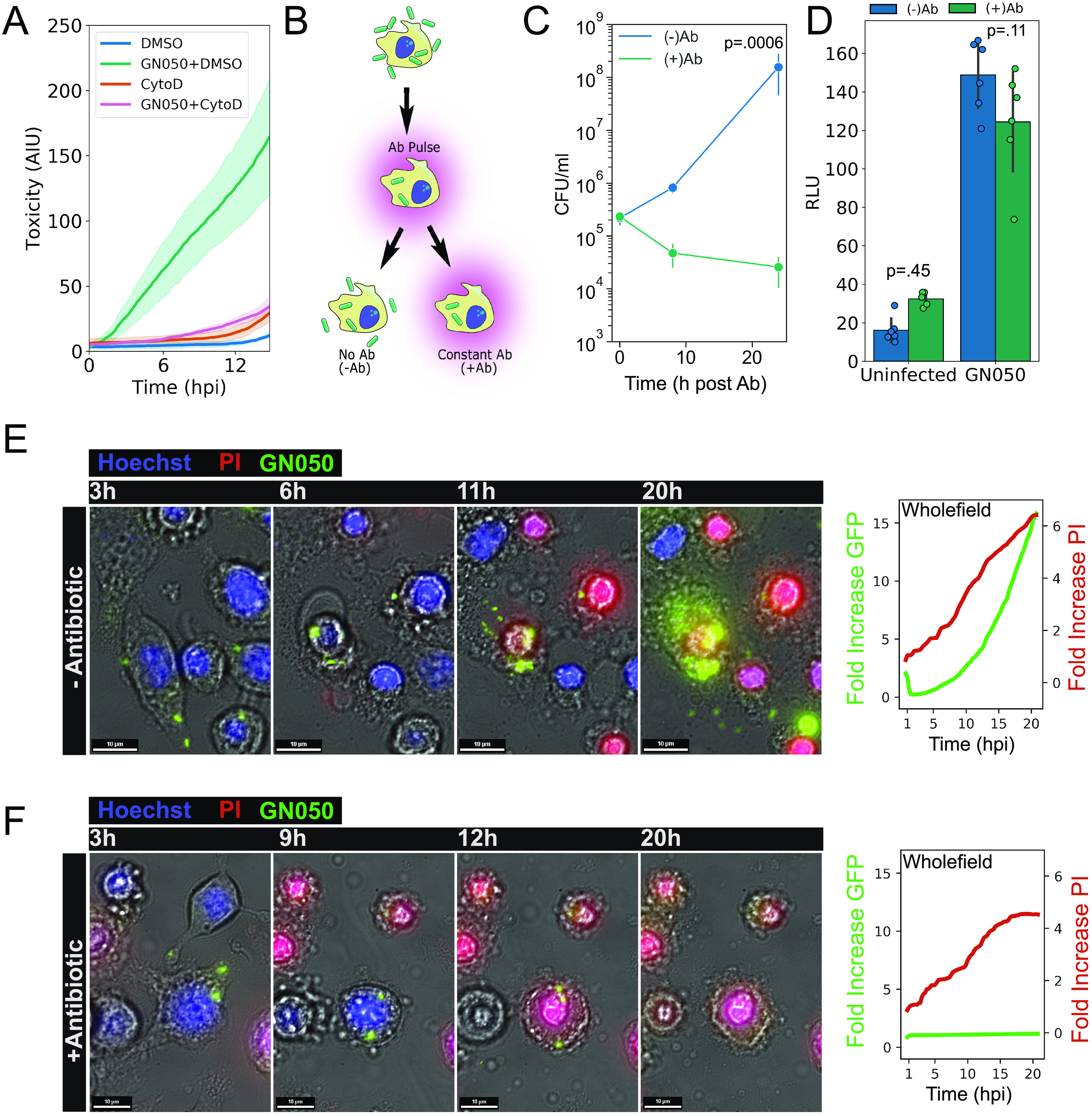
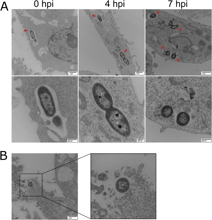
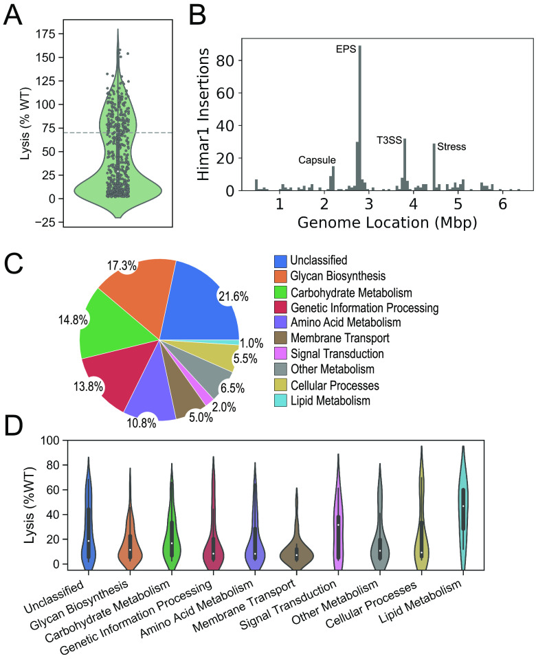
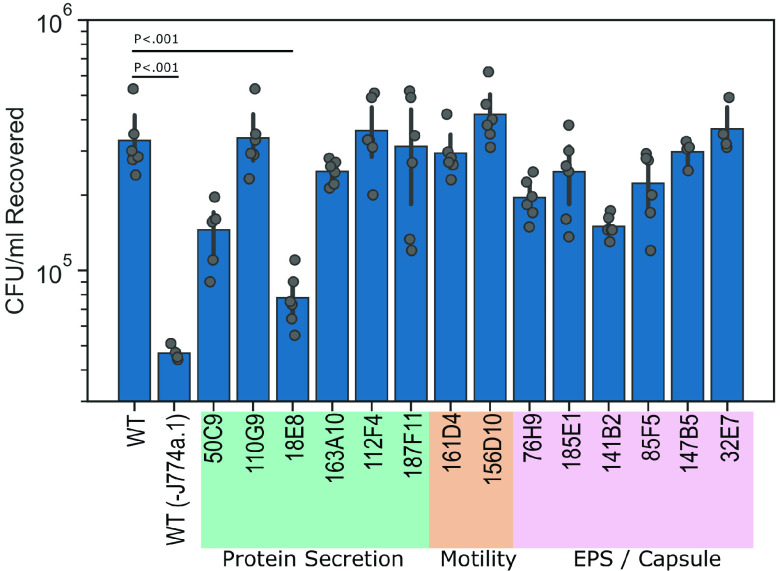
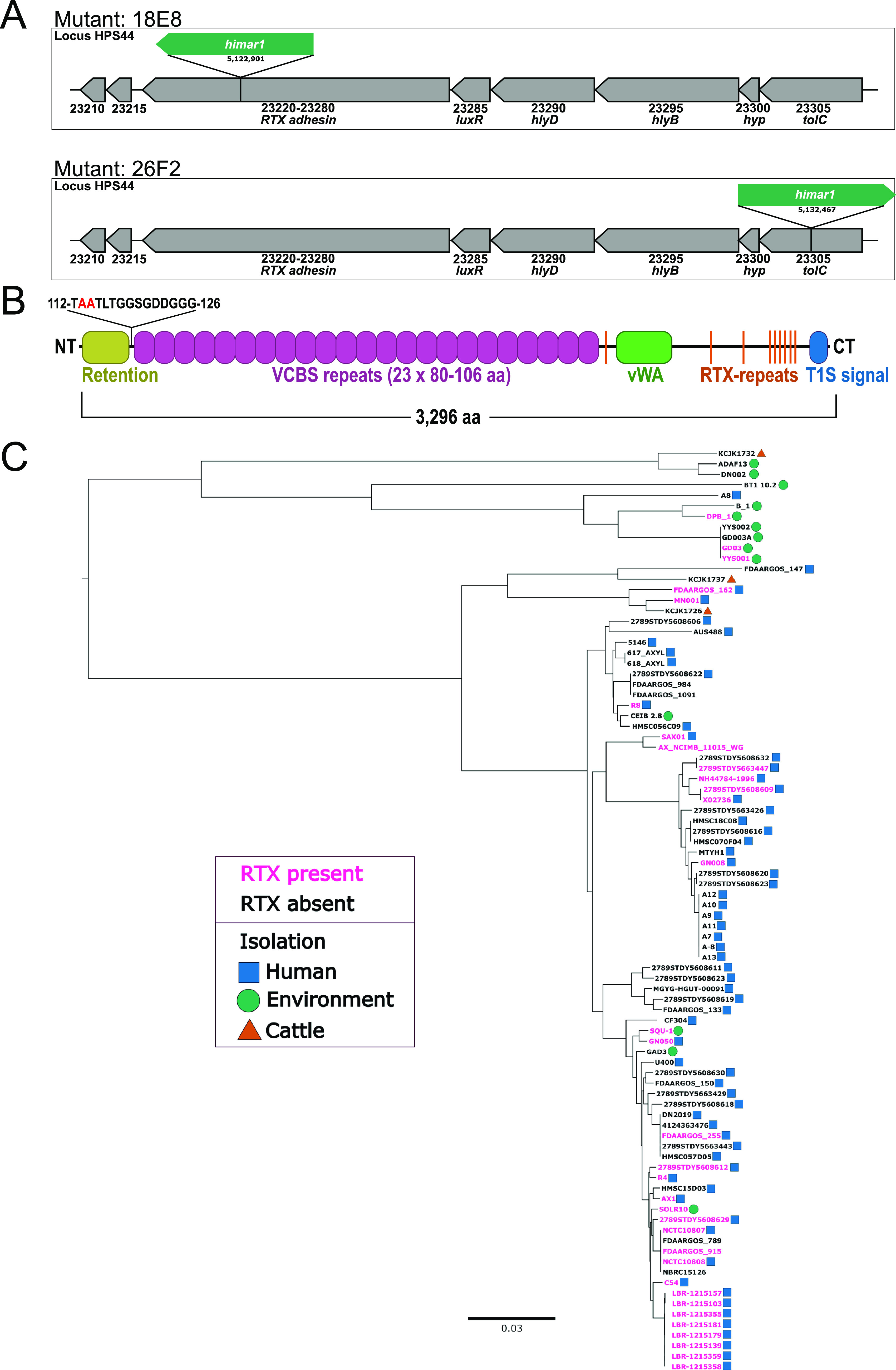
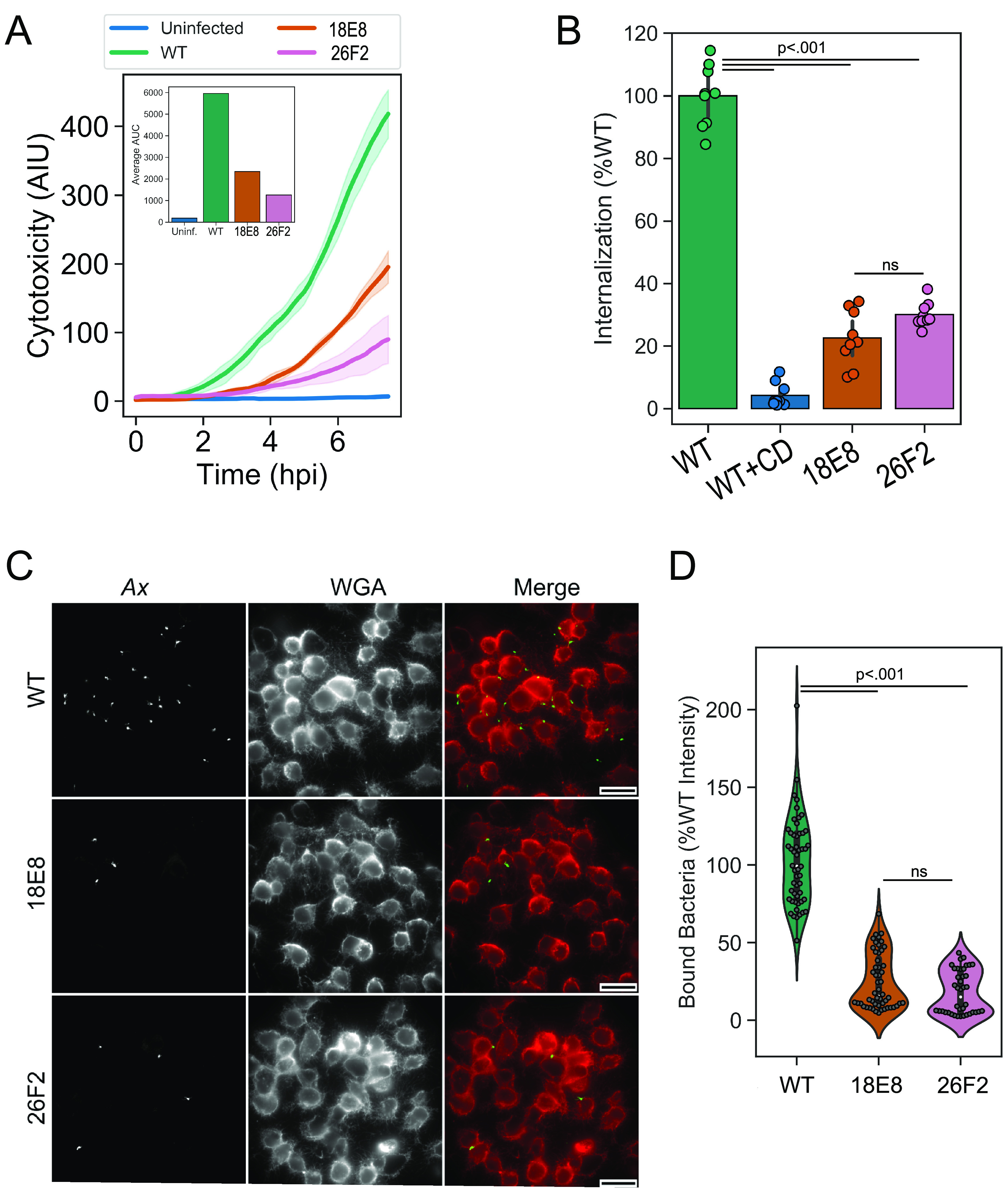
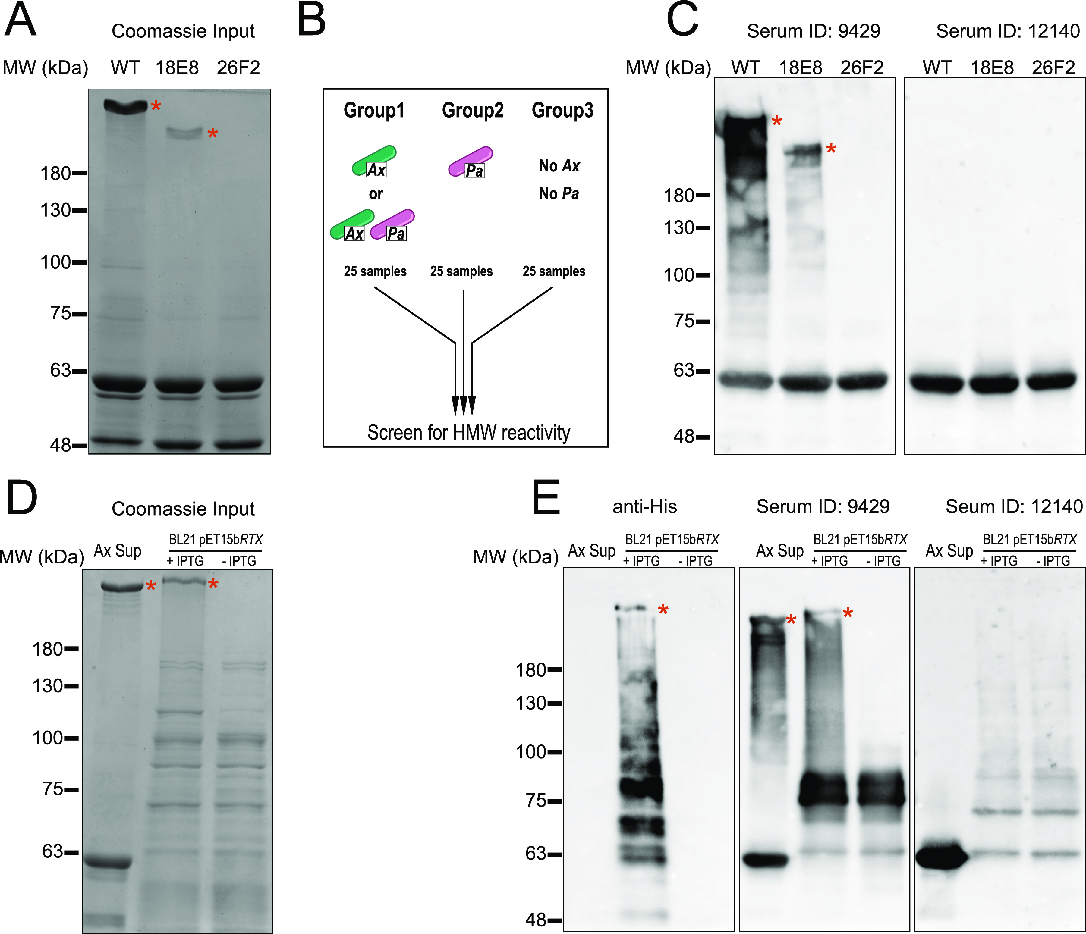
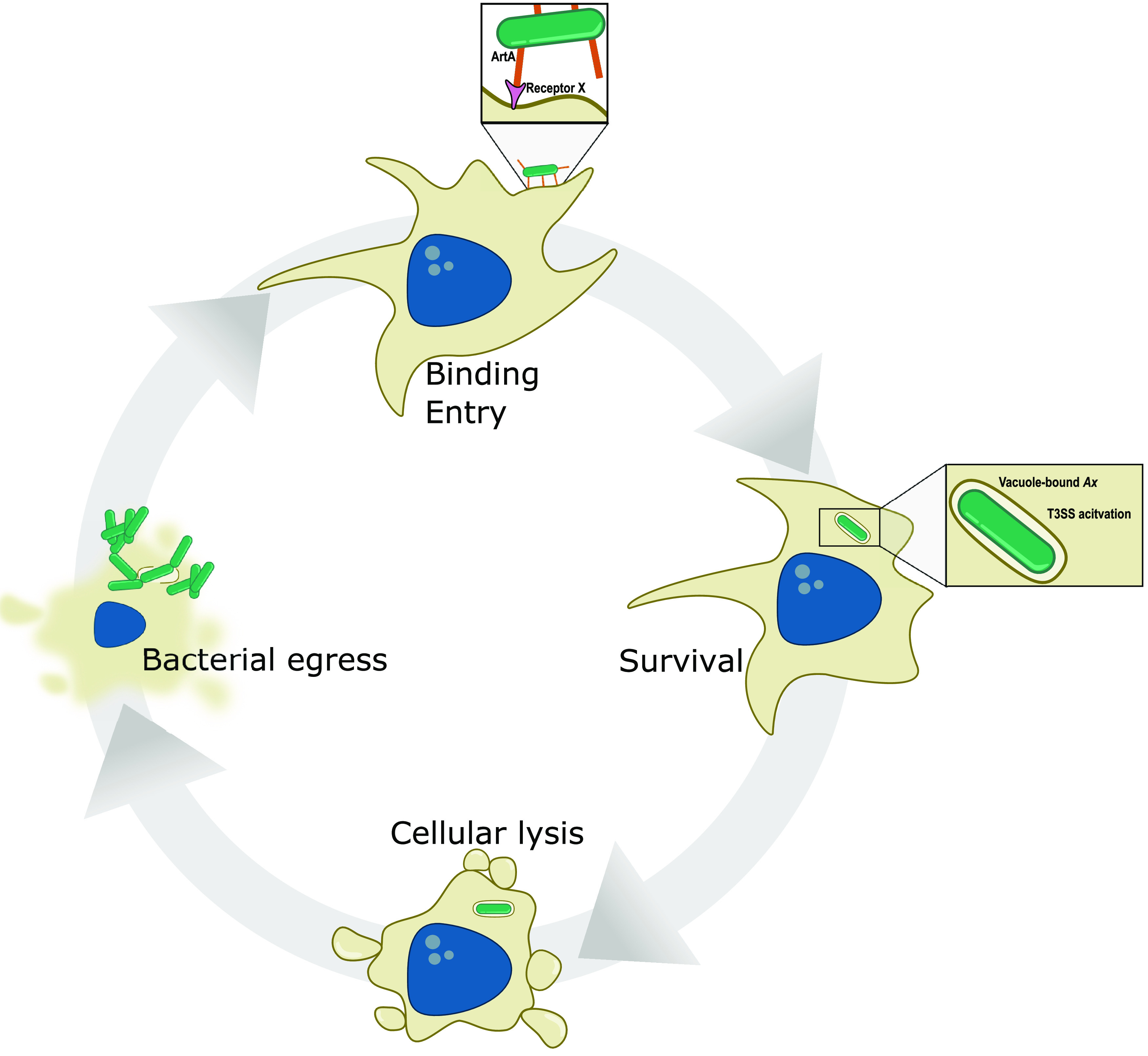
Achromobacter xylosoxidans is an opportunistic pathogen implicated in a wide variety of human infections including the ability to colonize the lungs of cystic fibrosis (CF) patients. The role of A. xylosoxidans in human pathology remains controversial due to the lack of optimized in vitro and in vivo model systems to identify and test bacterial gene products that promote a pathological response. We have previously identified macrophages as a target host cell for A. xylosoxidans-induced cytotoxicity. By optimizing our macrophage infection model, we determined that A. xylosoxidans enters macrophages and can reside within a membrane bound vacuole for extended periods of time. Intracellular replication appears limited with cellular lysis preceding an enhanced, mainly extracellular replication cycle. Using our optimized in vitro model system along with transposon mutagenesis, we identified 163 genes that contribute to macrophage cytotoxicity. From this list, we characterized a giant RTX adhesin encoded downstream of a type one secretion system (T1SS) that mediates bacterial binding and entry into host macrophages, an important first step toward cellular toxicity and inflammation. The RTX adhesin is encoded by other human isolates and is recognized by antibodies present in serum isolated from CF patients colonized by A. xylosoxidans, indicating this virulence factor is produced and deployed in vivo. This study represents the first characterization of A. xylosoxidans replication during infection and identifies a variety of genes that may be linked to virulence and human pathology. IMPORTANCE Patients affected by CF develop chronic bacterial infections characterized by inflammatory exacerbations and tissue damage. Advancements in sequencing technologies have broadened the list of opportunistic pathogens colonizing the CF lung. A. xylosoxidans is increasingly recognized as an opportunistic pathogen in CF, yet our understanding of the bacterium as a contributor to human disease is limited. Genomic studies have identified potential virulence determinants in A. xylosoxidans isolates, but few have been mechanistically studied. Using our optimized in vitro cell model, we identified and characterized a bacterial adhesin that mediates binding and uptake by host macrophages leading to cytotoxicity. A subset of serum samples from CF patients contains antibodies that recognize the RTX adhesion, suggesting, for the first time, that this virulence determinant is produced in vivo. This work furthers our understanding of A. xylosoxidans virulence factors at a mechanistic level.

Keywords: Achromobacter; RTX; cystic fibrosis; cytotoxicity; pathogenesis.


Conflict of interest statement

The authors declare no conflict of interest.

Figures

FIG 1
FIG 1
Growth of Ax GN050 during macrophage infection. (A) J774a.1 cells were pretreated with cytochalasin D or an equivalent volume of DMSO prior to GN050 infection (MOI: 1). PI was included over the course of infection and was quantified using CellProfiler software as a readout for host cell toxicity. The solid line is the mean, and the shaded areas are the error from four fields of view. (B) Infection schematic for subsequent assays in the figure. J774a.1 cells were incubated with GN050 for 30 min to allow for phagocytosis followed by treatment with gentamicin and polymyxin B to kill extracellular bacteria. Infection medium was replaced with fresh medium (−Ab) to allow intracellular and extracellular bacterial growth or antibiotic containing medium (+Ab) to select for intracellular bacteria only. (C) Quantification of GN050 (MOI: 1) during J774a.1 infection under −Ab or +Ab conditions (P = 0.0006 at 24 hpi). Data points are the mean of three independent infections (n = 6). (D) Cytotoxicity of GN050-infected J774a.1 cells (MOI: 1) under −Ab or +Ab conditions as determined by AK release (n = 6). Data were assessed by two-way ANOVA followed by Tukey’s post hoc analysis. (E and F) Live imaging analysis of GN050-infected J774a.1 cells (MOI: 1) from 1 to 20 hpi under −Ab (E) or +Ab (F) conditions. Stills from the live imaging were cropped and merged (left). Quantification of host cytotoxicity (fold increase PI) and GN050 proliferation (fold increase GFP) from full capture fields are on the right. Images and quantification are from a single representative infection that was performed at least on three separate occasions. Scale = 10 μm.
FIG 2
FIG 2
Subcellular localization of Ax GN050 during macrophage infection by transmission electron microscopy. (A) J774a.1 macrophages were infected with GN050 (MOI: 1) for 30 min and washed, and the medium was replaced with antibiotic-containing medium for 30 min. Antibiotic medium was replaced with fresh medium and samples were taken at 4 hpi and 7 hpi. An initial time point (0 hpi) was taken at 5 min postinoculation. (A) Vacuole-based GN050 (red arrows) were observed at all time points at low (top) and high (bottom) magnification images. (B) Cellular lysis and bacterial egress from a GN050-infected J774a.1 cell at 7 hpi. Representative images are from two independent infections.
FIG 3
FIG 3
Transposon mutagenesis of Ax strain GN050 (A) Cytotoxicity of J774a.1 macrophages infected with GN050 mutants (MOI: 10 for 8 h) identified in the initial CV-based screen was reassessed in a secondary screen using an AK release assay (n = 2). GN050 mutants equal to or below 70% of WT cytotoxicity (dashed line) were subjected to further analysis. (B) Identified himar1 insertions sites across the Ax genome when individual insertion events from each mutant were compiled. Each bar represents a 64.5-kb segment of the GN050 genome. (C) Classification of functional pathways involved in macrophage cytotoxicity. Amino acid sequences of ORFs containing himar1 insertions were annotated using KEGG databases. (D) Data sets from panels A and C were merged to visualize the relationship between a cytotoxicity defect of a mutant and the disrupted pathway.
FIG 4
FIG 4
Cell association screen used to identify GN050 mutants defective for host cell binding or entry. GN050 mutant candidates were selected based on disruption of genes predicted to be involved in the production or transport of extracellular substrates. Mutants were incubated with J774a.1 macrophages (MOI: 1) for 30 min followed by two washes to remove extracellular, unbound bacteria. Wells were emulsified and the remaining bacteria were enumerated. To control for background adherence, WT GN050 were incubated without J774a.1 macrophages, WT(-J774a.1). Data bars are the mean, and the error is ± 1 SD. Data points of n = 4 to 6 from 2 to 3 biological replicates were assessed by one-way ANOVA followed by Tukey’s post hoc analysis.
FIG 5
FIG 5
Genomic analysis of the T1SS/artA locus. (A) T1SS/artA coding region and location of the transposon insertion in mutants 18E8 and 26F2. Locus tag HPS44_23305, tolC OM protein. 23300, hypothetical. 23295, hlyB permease/ATPase. 23290, hlyD periplasmic adaptor. 23285, luxR transcriptional regulator. 23220-23280, artA RTX adhesin. 23215, transglutaminase. 23210, EAL domain-containing protein. (B) Domain map of the RTX aa sequence determined by InterProScan. Putative N-terminal cleavage site is highlighted in red. (C) Maximum likelihood phylogenetic tree (midpoint root) of 88 Ax isolates based on 77 single-copy core genes. Presence (pink) or absence (black) of full or partial ArtA in Ax genomes along with isolation source is denoted. Blue squares, human isolation. Green circles, environmental isolation. Orange triangles, cattle isolation.
FIG 6
FIG 6
Binding and internalization defects contribute to cytotoxicity defects in 18E8 and 26F2. (A) Live imaging of J774a.1 infected with GN050 or derived strains (MOI: 5) for 8 h. Solid lines are the mean intensity of PI incorporation, or arbitrary intensity units (AIU), into damaged cells from n = 4 fields of view (10× objective) and the shaded region is ± 1 SD. The inset is the average area under the curve (AUC) for each group. (B) Internalization of GN050 and derived strains (MOI: 1) after 30 min of phagocytosis by J774a.1 cells. Cells were either pretreated with cytochalasin D (CD) or an equal volume of DMSO. Internalization was normalized relative to WT over the course of three independent experiments (n = 9). Data bars are the mean, and the error is ± 1 SD (C) Representative IF microscopy images of J774a.1 cells incubated with WT GN050 or derived strains (MOI: 100). Cells were fixed and labeled with polyclonal antibody recognizing GN050 outer membrane proteins (Ax) and wheat germ agglutinin (WGA) to visualize the macrophage membrane. Scale = 25 μm. (D) Quantification of Ax signal intensity from 40 to 60 fields of view (10× objective) over 2 to 3 independent experiments and values were normalized to WT binding levels. Data were assessed by one-way ANOVA followed by Tukey’s post hoc analysis.
FIG 7
FIG 7
ArtA is released into cultured supernatants and serum from CF patients contain antibodies recognizing ArtA. (A) Coomassie stained SDS-PAGE gel of concentrated culture supernatants from WT GN050, 18E8 and 26F2. (B) Schematic groups of CF serum used to screen for reactivity against ArtA. Group 1, colonization with Ax or Ax and P. aeruginosa (Pa). Group 2, colonization with Pa but not Ax. Group 3, no history of colonization with Pa or Ax. (C) Reactivity of group one serum from CF patients with the input antigen from panel A. (D) ArtA production in E. coli BL21 visualized by Coomassie after SDS-PAGE. WT GN050 precipitated supernatant was used as a positive control for the presence of the HMW product (lane 1). ArtA production under induced (lane 2) or uninduced (lane 3) conditions were analyzed. (E) Using the antigen input in panel C, immunoblotting using anti-His, serum 9429 or serum 12140 was used to detect reactivity to the RTX adhesin. In panels A to E, the presence of multiple forms of the GN050 RTX adhesin is marked by a red asterisk. Data are representative of similar experiments performed on multiple occasions.
FIG 8
FIG 8
Proposed model for GN050 toxicity in macrophages. The GN050 infection cycle begins by binding to the host cell which is enhanced by the GN050 RTX adhesin ArtA binding to a putative receptor. After bacterial internalization, GN050 remains in a vacuole where T3SS genes and a putative toxin, axoU, are transcriptionally activated (36). Survival of vacuole-based GN050 for up to 9 h is followed by host cell membrane blebbing and rupture. GN050 is then released into the extracellular space where growth occurs followed by subsequent internalization events.

Similar articles

Cited by

References

    1. Yabuuchi E, Oyama A. 1971. Achromobacter xylosoxidans n. sp. from human ear discharge. Jpn J Microbiol 15:477–481. doi:10.1111/j.1348-0421.1971.tb00607.x. - DOI - PubMed
    1. Neidhöfer C, Berens C, Parčina M. 2022. An 18-year dataset on the clinical incidence and MICs to antibiotics of Achromobacter spp. (labeled biochemically or by MAL-DI-TOF MS as A. xylosoxidans), largely in patient groups other than those with CF. Antibiotics 11:311. doi:10.3390/antibiotics11030311. - DOI - PMC - PubMed
    1. Emerson J, McNamara S, Buccat AM, Worrell K, Burns JL. 2010. Changes in cystic fibrosis sputum microbiology in the United States between 1995 and 2008. Pediatr Pulmonol 45:363–370. doi:10.1002/ppul.21198. - DOI - PubMed
    1. Spilker T, Vandamme P, LiPuma JJ. 2013. Identification and distribution of Achromobacter species in cystic fibrosis. J Cyst Fibros 12:298–301. doi:10.1016/j.jcf.2012.10.002. - DOI - PubMed
    1. Pereira RHV, Leão RS, Carvalho-Assef AP, Albano RM, Rodrigues ERA, Firmida MC, Folescu TW, Plotkowski MC, Bernardo VG, Marques EA. 2017. Patterns of virulence factor expression and antimicrobial resistance in Achromobacter xylosoxidans and Achromobacter ruhlandii isolates from patients with cystic fibrosis. Epidemiol Infect 145:600–606. doi:10.1017/S0950268816002624. - DOI - PMC - PubMed

Publication types

MeSH terms