

Review
doi: 10.1259/bjr.20211125.
Epub 2022 Aug 3.
A review of inferior vena cava filters
Affiliations
- PMID: 35856774
- PMCID: PMC10997026
- DOI: 10.1259/bjr.20211125
Item in Clipboard
Review
A review of inferior vena cava filters
Br J Radiol.
.
Abstract
The care of patients with venous thromboembolism (VTE) is delivered via a multidisciplinary team. The primary treatment for VTE is anticoagulation; however, placement of filter devices in the inferior vena cava (IVC) to prevent embolisation of deep venous thrombosis (DVT) is a well-established secondary treatment option. Many controversies remain regarding utilisation and management of filters.
Figures
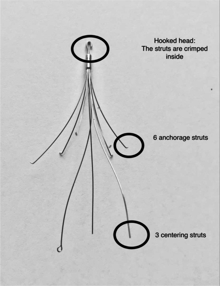
The ALN Filter (Implants Chirurgicaux, France) is our current filter of
choice.
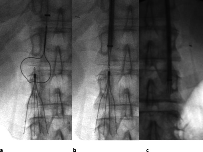
Celect® (Cook) IVC filter removed with retrieval snare set in a
31-year-old male with a history of PE, testicular cancer and retroperitoneal
lymph node dissection. (a) After first performing a venogram to ensure no
thrombus is present in the filter, the looped snare is manipulated down over
the hook. (b) The snare has been tightened around the hook and (c) the
sheath is pushed down over the filter while holding the snared filter firmly
until the filter is within the sheath. The filter is then removed through
the sheath. IVC, inferior vena cava; PE, pulmonary embolism.
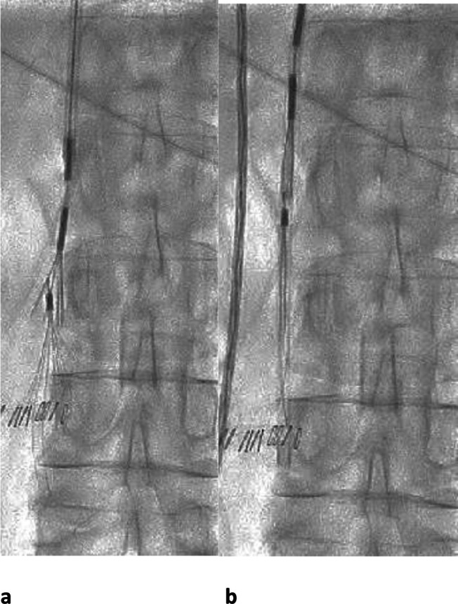
ALN IVC filter retrieval in a 77-year-old lady who had a DVT and needed
interruption of anticoagulation for a neurosurgical procedure. (a) 6 weeks
after the surgery, the ALN retrieval device was used for retrieval (grasping
device with curve on the end of the delivery sheath to facilitate removal of
filters tips close to the caval wall). The grasper of the retrieval device
is seen directed over the hook of the filter. (b) Once the grasping device
has engaged the filter tip, it is closed around it and the sheath advanced
over the filter, which is then withdrawn. IVC, inferior vena cava; DVT, deep
venous thrombosis.
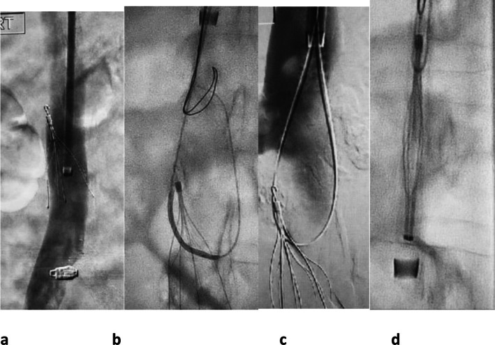
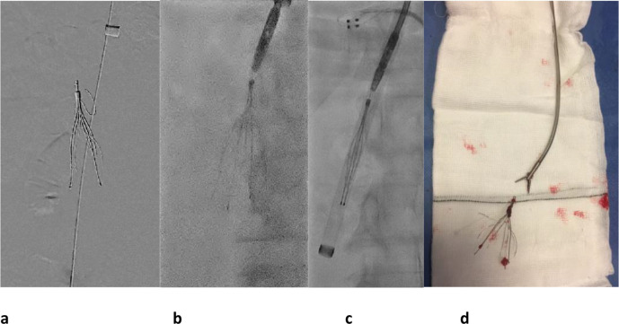
Example of a difficult retrieval. Initial standard retrieval methods had
failed as (a) the filter had tilted against the wall of the IVC and
endothelialised. (b) Using a long 16 Fr sheath and a Rim catheter
(AngioDynamics, New York) , a 300 cm x 0.014 inch pilot wire was looped
through the struts of the filter. (c) The 0.014 inch guidewire was then
snared using a filter removal loop snare. The guidewire was brought out
through the sheath to the skin so that a long loop of guidewire is present
from the skin to the tip of the filter. (d) The filter was straightened by
pulling both ends of the guidewire and the sheath was advanced over the hook
and the filter removed. IVC, inferior vena cava.
51-year-old lady with unprovoked above knee DVT, saddle pulmonary embolus,
and intracranial bleed had an IVC filter inserted. Multiple attempts were
made to remove the filter with standard snare techniques, but the hook of
the filter was embedded in the anterior wall. (a) Attempts at removing the
filter caused an arm strut to bend cranially. (b) The patient had a 16 Fr
long sheath placed through which an ENT forceps, which was manually curved
before insertion, was manipulated on to the hook at the top of the filter
and the hook grasped. (c) While keeping the forceps closed around the hook
of the filter, the sheath was manipulated down over the filter and the
filter removed. (d) The filter and ENT forceps are shown ex vivo. IVC,
inferior vena cava; DVT, deep venous thrombosis.
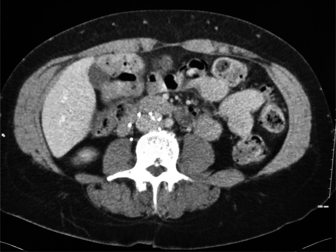
Axial CT demonstrates IVC filter wall penetration of an anchoring strut
toward the duodenum on the right. IVC, inferior vena cava.
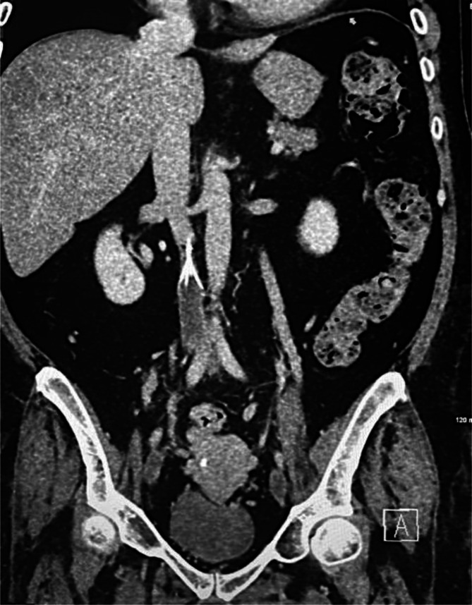
Coronal CT image demonstrates IVC thrombus below the filter. IVC, inferior
vena cava.
References
-
- Liu Y, Lu H, Bai H, Liu Q, Chen R. Effect of inferior vena cava filters on pulmonary embolism-related mortality and major complications: a systematic review and meta-analysis of randomized controlled trials. J Vasc Surg Venous Lymphat Disord 2021; 9: 792–800. doi: 10.1016/j.jvsv.2021.02.008 - DOI - PubMed
Publication types
MeSH terms
LinkOut - more resources
Full Text Sources
Medical
