

Basic Fibroblast Growth Factor Induces Cholinergic Differentiation of Tonsil-Derived Mesenchymal Stem Cells
- PMID: 35857260
- PMCID: PMC9478007
- DOI: 10.1007/s13770-022-00474-0
Basic Fibroblast Growth Factor Induces Cholinergic Differentiation of Tonsil-Derived Mesenchymal Stem Cells
Abstract
Background: Mesenchymal stem cells (MSCs) are considered a potential tool for regenerating damaged tissues due to their great multipotency into various cell types. Here, we attempted to find the appropriate conditions for neuronal differentiation of tonsil-derived MSCs (TMSCs) and expand the potential application of TMSCs for treating neurological diseases.
Methods: The TMSCs were differentiated in DMEM/F-12 (Dulbecco's Modified Eagle Medium/Nutrient Mixture F-12) supplemented with various neurotrophic factors for 7-28 days to determine the optimal neuronal differentiation condition for the TMSCs. The morphologies as well as the levels of the neural markers and neurotransmitters were assessed to determine neuronal differentiation potentials and the neuronal lineages of the differentiated TMSCs.
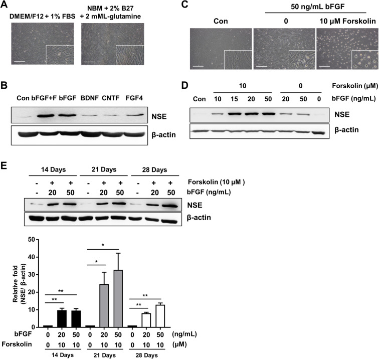
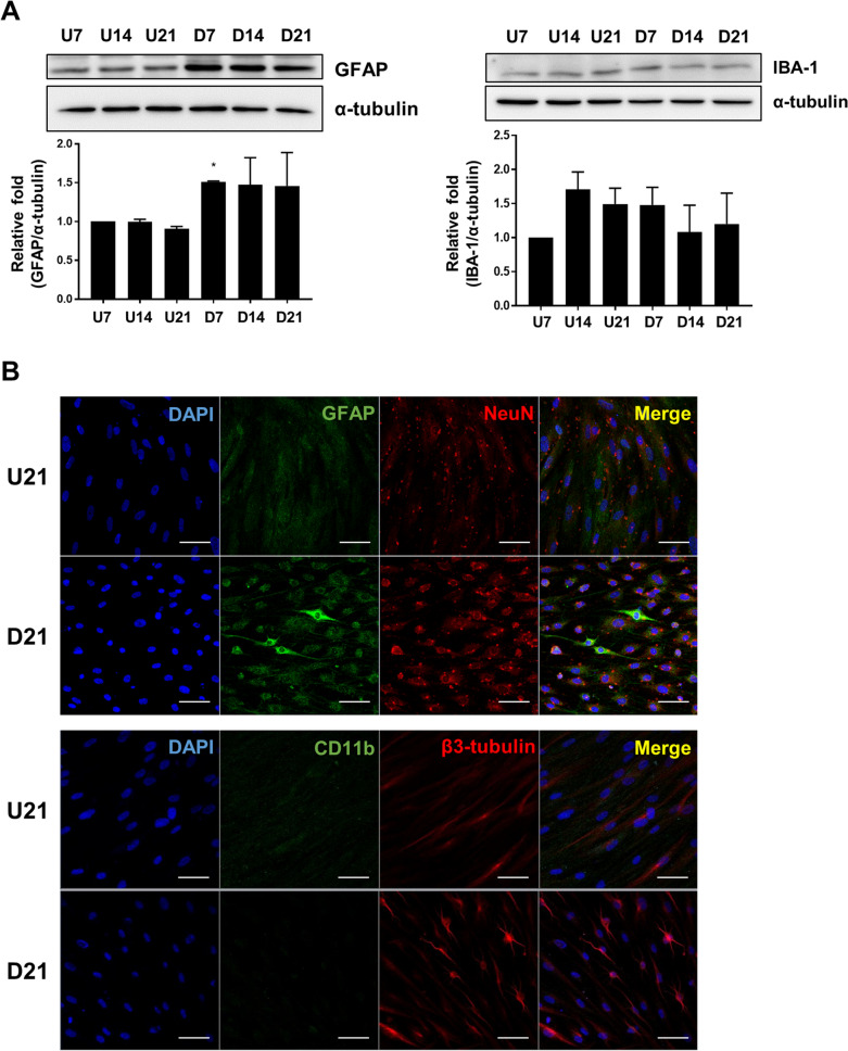
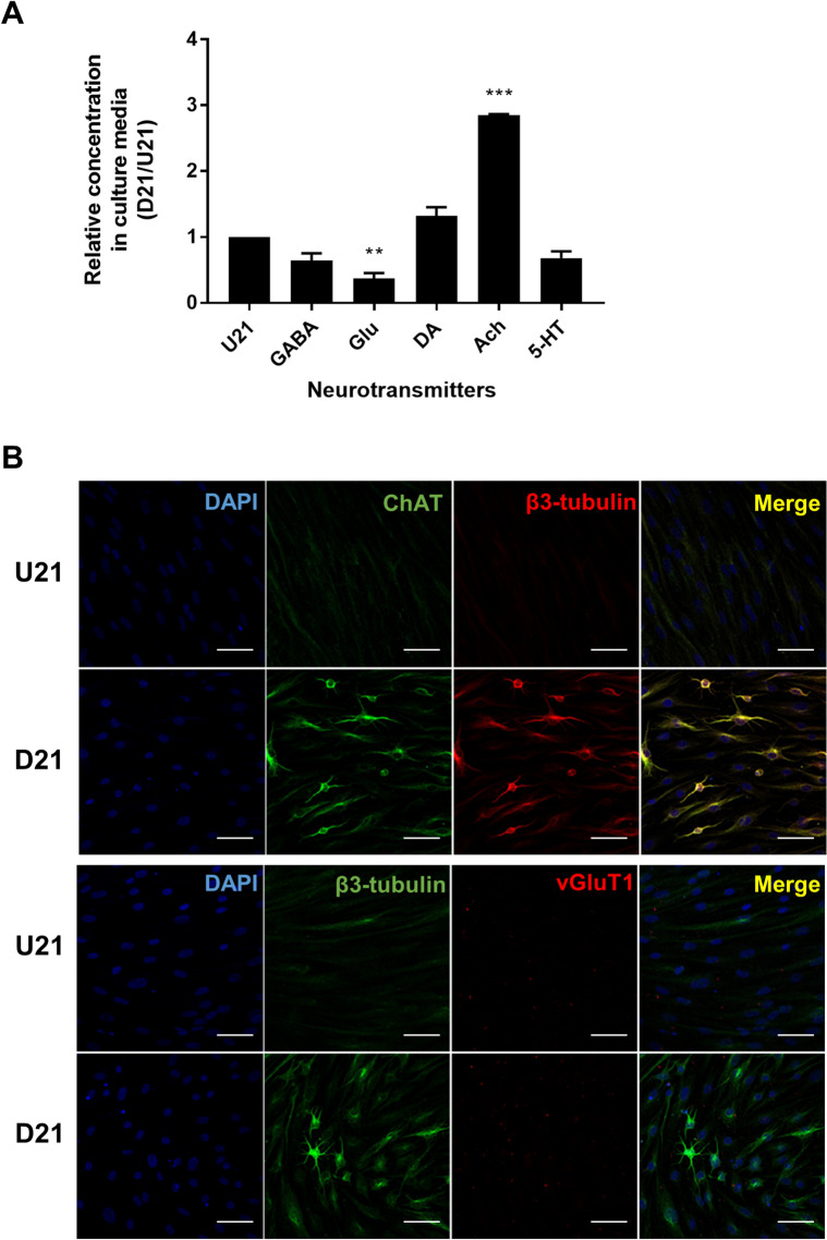
Results: Our initial study demonstrated that DMEM/F12 supplemented with 50 ng/mL basic fibroblast growth factor with 10 μM forskolin was the optimal condition for neuronal differentiation for the TMSCs. TMSCs had higher protein expression of neuronal markers, including neuron-specific enolase (NSE), GAP43, postsynaptic density protein 95 (PSD95), and synaptosomal-associated protein of 25 kDa (SNAP25) compared to the undifferentiated TMSCs. Immunofluorescence staining also validated the increased mature neuron markers, NeuN and synaptophysin, in the differentiated TMSCs. The expression of glial fibrillar acidic protein and ionized calcium-binding adaptor molecule 1 the markers of astrocytes and microglia, were also slightly increased. Additionally, the differentiated TMSCs released a significantly higher level of acetylcholine, the cholinergic neurotransmitter, as analyzed by the liquid chromatography-tandem mass spectrometry and showed an enhanced choline acetyltransferase immunoreactivity compared to the undifferentiated cells.
Conclusion: Our study suggests that the optimized condition favors the TMSCs to differentiate into cholinergic neuron-like phenotype, which could be used as a possible therapeutic tool in treating certain neurological disorders such as Alzheimer's disease.
Keywords: Basic fibroblast growth factor; Neuronal differentiation; Tonsil-derived mesenchymal stem cells.
© 2022. Korean Tissue Engineering and Regenerative Medicine Society.
Conflict of interest statement
The authors declare no conflict of interest.
Figures

Similar articles
-
Optimization of Microenvironments Inducing Differentiation of Tonsil-Derived Mesenchymal Stem Cells into Endothelial Cell-Like Cells.Tissue Eng Regen Med. 2019 Oct 30;16(6):631-643. doi: 10.1007/s13770-019-00221-y. eCollection 2019 Dec. Tissue Eng Regen Med. 2019. PMID: 31824825 Free PMC article.
-
A transcriptomic analysis of serial-cultured, tonsil-derived mesenchymal stem cells reveals decreased integrin α3 protein as a potential biomarker of senescent cells.Stem Cell Res Ther. 2020 Aug 17;11(1):359. doi: 10.1186/s13287-020-01860-y. Stem Cell Res Ther. 2020. PMID: 32807231 Free PMC article.
-
A Novel Method to Differentiate Tonsil-Derived Mesenchymal Stem Cells In Vitro into Estrogen-Secreting Cells.Tissue Eng Regen Med. 2021 Apr;18(2):253-264. doi: 10.1007/s13770-020-00307-y. Epub 2020 Oct 28. Tissue Eng Regen Med. 2021. PMID: 33113109 Free PMC article.
-
Therapeutic Effect of IL1β Priming Tonsil Derived-Mesenchymal Stem Cells in Osteoporosis.Tissue Eng Regen Med. 2021 Oct;18(5):851-862. doi: 10.1007/s13770-021-00350-3. Epub 2021 Jun 11. Tissue Eng Regen Med. 2021. PMID: 34115339 Free PMC article.
-
Application of Tonsil-Derived Mesenchymal Stem Cells in Tissue Regeneration: Concise Review.Stem Cells. 2019 Oct;37(10):1252-1260. doi: 10.1002/stem.3058. Epub 2019 Jul 25. Stem Cells. 2019. PMID: 31287931 Free PMC article. Review.
Cited by
-
Stepwise differentiation of airway epithelial cells from human tonsil-derived mesenchymal stem cells.Stem Cell Res Ther. 2025 Jul 7;16(1):354. doi: 10.1186/s13287-025-04397-0. Stem Cell Res Ther. 2025. PMID: 40624536 Free PMC article.
-
Impact of 17β-Estradiol on the Shape, Survival, Osteogenic Transformation, and mRNA Expression of Gingiva-Derived Stem Cell Spheroids.Medicina (Kaunas). 2023 Dec 28;60(1):60. doi: 10.3390/medicina60010060. Medicina (Kaunas). 2023. PMID: 38256321 Free PMC article.
References
Publication types
MeSH terms
Substances
LinkOut - more resources
Full Text Sources
Other Literature Sources
Miscellaneous