. 2022 Jul 15;8(28):eabn3326.
doi: 10.1126/sciadv.abn3326. Epub 2022 Jul 13.

Neuronal activity induces glucosylceramide that is secreted via exosomes for lysosomal degradation in glia

Affiliations

Neuronal activity induces glucosylceramide that is secreted via exosomes for lysosomal degradation in glia

Liping Wang et al. Sci Adv. .

Abstract

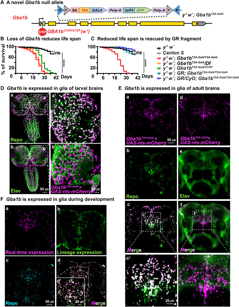
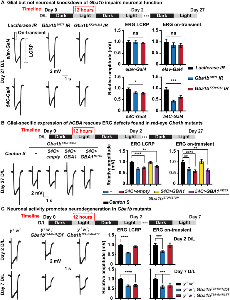
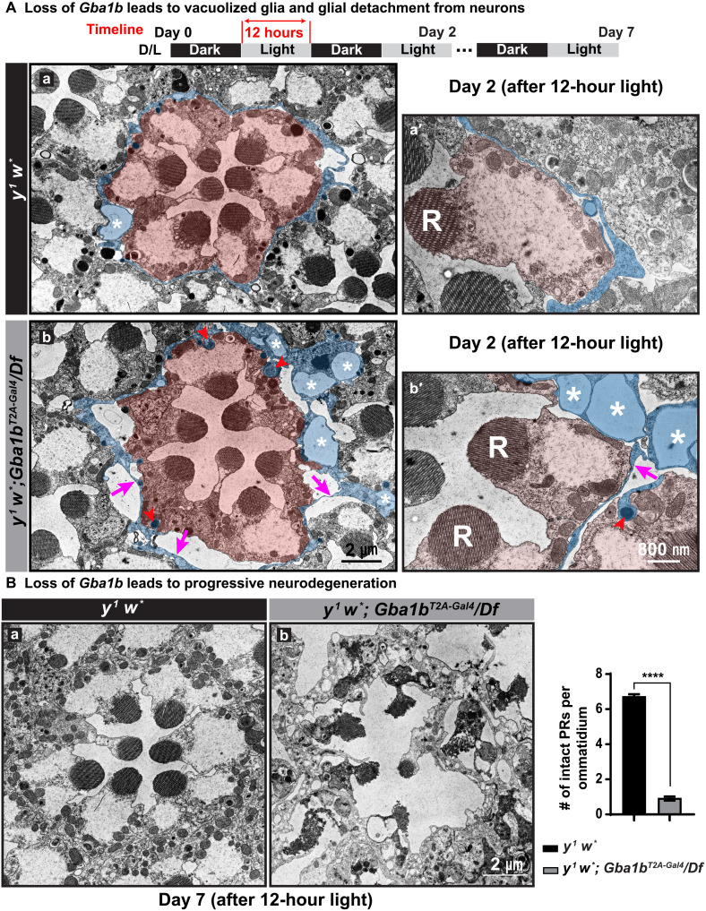
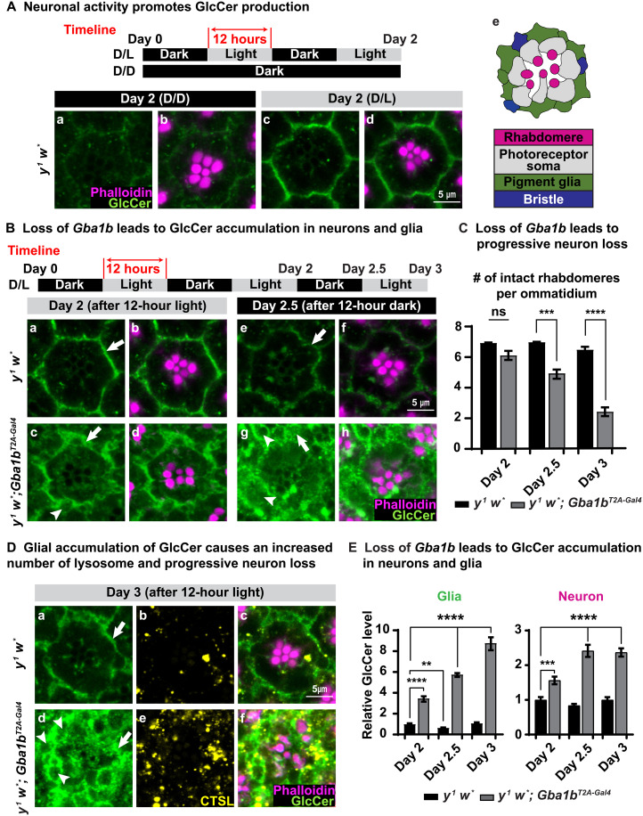
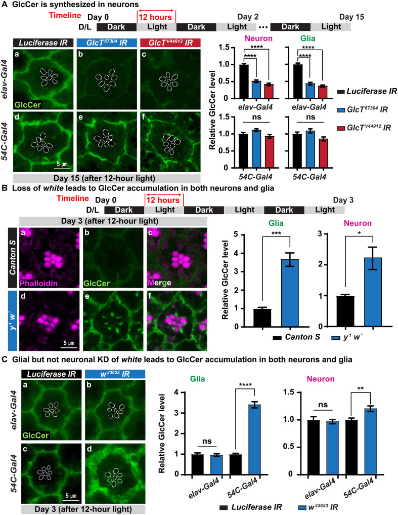
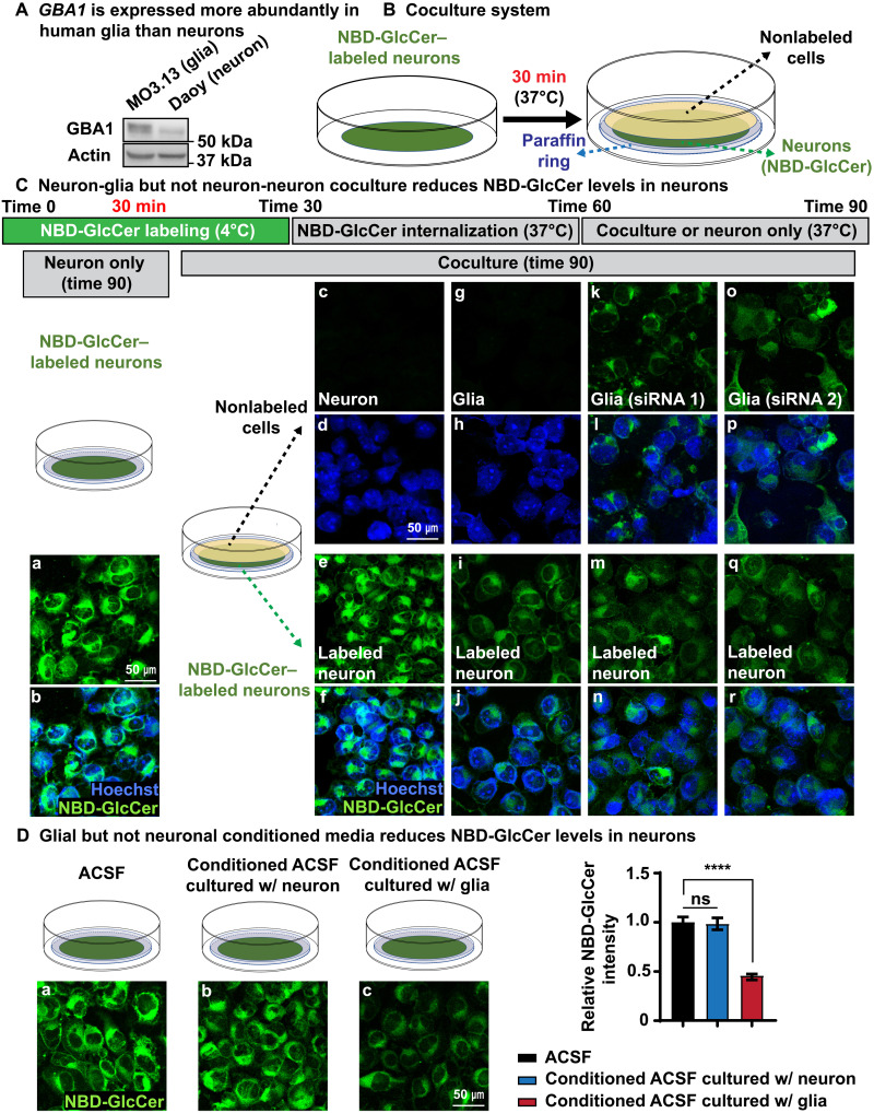
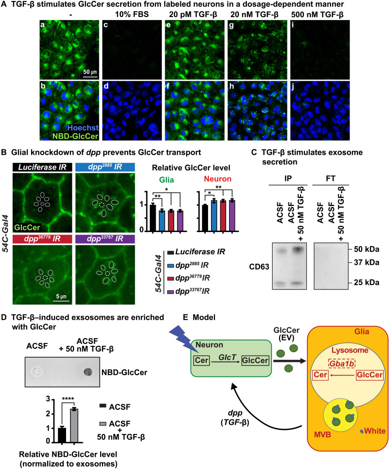
Recessive variants in GBA1 cause Gaucher disease, a prevalent form of lysosome storage disease. GBA1 encodes a lysosomal enzyme that hydrolyzes glucosylceramide (GlcCer) into glucose and ceramide. Its loss causes lysosomal dysfunction and increased levels of GlcCer. We generated a null allele of the Drosophila ortholog Gba1b by inserting the Gal4 using CRISPR-Cas9. Here, we show that Gba1b is expressed in glia but not in neurons. Glial-specific knockdown recapitulates the defects found in Gba1b mutants, and these can be rescued by glial expression of human GBA1. We show that GlcCer is synthesized upon neuronal activity, and it is transported from neurons to glia through exosomes. Furthermore, we found that glial TGF-β/BMP induces the transfer of GlcCer from neurons to glia and that the White protein, an ABCG transporter, promotes GlcCer trafficking to glial lysosomes for degradation.


Figures

Fig. 1.
Fig. 1.. Gba1b is expressed in glia.
(A) y1 w*; Gba1bT2A-Gal/T2A-Gal4: A construct containing attp-FRT-SA-T2A-GAL4-PolyA-3xP3GFP-FRT-attp was inserted in Gba1b using CRIMIC technology (32). This insertion creates a null allele as shown in fig. S1. It also leads to the production of GAL4 protein in the same temporal and spatial expression pattern as Gba1b. (B and C) Reduced life span of flies that lack Gba1b: These include (i) y1 w*; Gba1bT2A-Gal4/T2A-Gal4, (ii) y1 w*; Gba1bT2A-Gal4/Df, and (iii) y1 w*; Gba1bT2A-Gal/STOP (this is a red-eyed fly due to the presence of the Gba1b STOP allele). The Gba1bSTOP/STOP allele contains an early stop codon and an insertion of the mini-white+ gene (22). Reduced life span can be fully rescued by one or two copies of a genomic fragment (GR) that contains the Gba1b gene and the white+ gene (N > 200). ****P < 0.0001. (D and E) Gba1b is expressed in glia in larval and adult brains. The GAL4 protein produced by the y1 w*; Gba1bT2A-Gal4 insertion is used to drive the nls::mCherry protein (magenta). On the basis of costaining with Repo (green) present in the nuclei of glia, Gba1b is only expressed in glia. Moreover, there is no obvious colocalization between Elav (green), which marks neuronal nuclei, and nls::mCherry, which reports Gba1b expression, indicating that Gba1b is not expressed in neurons (N = 3). (F) Magenta and green signals represent real-time and lineage expression of Gba1b, respectively (see fig. S1, B and C). Again, Gba1b is only expressed in glia. Repo (canyon) labels the glial nuclei (N = 3).
Fig. 2.
Fig. 2.. Glial Gba1b is necessary and sufficient to support neuronal function.
(A) Gba1b is required in glia to support neuronal function. ERG recordings of flies of the indicated genotypes after 27 days of D/L cycles. Gba1b is specifically knocked down in neurons driven by elav-Gal4 (top) and in glia driven by 54C-Gal4 (bottom), respectively. Glial knockdown but not neuronal knockdown of Gba1b leads to reduction of LCRPs and on-transient. The ERG LCRP and on-transient amplitudes are quantified on the right. Error bars represent SEM (n ≥ 6); *P < 0.05 and ***P < 0.001. (B) Glial-specific expression of human GBA1 but not GBA1N370S rescues ERG phenotypes in Gba1b mutants. ERG recordings of flies of the indicated genotypes after 27 days of D/L cycles. Gba1bSTOP/STOP shows reduced ERG LCRP and on-transient, which can be fully rescued by glial expression of GBA1 driven by 54C-Gal4. The ERG LCRP and on-transient amplitudes are quantified on the right. Error bars represent SEM (n ≥ 6); **P < 0.01 and ****P < 0.0001. Flies that are tested in this experiment are red eyed because of the transgenes. (C) Neuronal activity promotes neurodegeneration in Gba1b mutants. ERG recordings of flies of the indicated genotypes after 2 and 7 days of D/L cycles. y1 w*; Gba1bΔTT is a partial loss-of-function allele with 40% remaining enzymatic activity (21). At day 2 (top), loss of Gba1b (y1 w*; Gba1bT2A-Gal4/Df) causes reduced ERG LCRP and on-transient amplitudes, whereas the partial loss-of-function allele (y1 w*; Gba1bT2A-Gal4/ΔTT) does not obviously affect the ERGs. At day 7 (bottom), both allelic combinations exhibit reduced LCRP and on-transient amplitudes. The data are quantified on the right. Error bars represent SEM (n ≥ 6); ***P < 0.001 and ****P < 0.0001. Flies that are tested in this experiment are phenotypically white.
Fig. 3.
Fig. 3.. Loss of Gba1b impairs glial morphology, which precedes neuronal loss.
(A) Loss of Gba1b leads to vacuolized glia. TEM images of fly retina of the indicated genotypes after 2 days of D/L cycles. Photoreceptor neurons are highlighted in orange, and pigment cells are highlighted in blue (a, a′, b, and b′). R, rhabdomeres. Red arrowheads point to lysosomes in pigment cells. y1 w*; Gba1bT2A-Gal4/Df null mutants exhibit an increased number of lysosomes in glia when compared to glia of y1 w* flies (b and b′). Magenta arrows indicate glial detachment, which is commonly seen in the retina of y1 w*; Gba1bT2A-Gal4/Df null mutants. Glial vacuoles are frequently seen in the retina of null mutant flies, and asterisks mark vacuoles in glia, which are rarely observed in y1 w* flies. (B) TEM images of fly retina of the indicated genotypes upon 7 days of D/L cycles. The overall morphology of the retina is severely affected in y1 w*; Gba1bT2A-Gal4/Df flies, whereas the retinas of y1 w* flies do not show obvious defects. The number of intact photoreceptors (PRs) per ommatidium is quantified on the right. Error bars represent SEM (n = 3); ****P < 0.0001. Flies that are tested in this experiment are phenotypically white.
Fig. 4.
Fig. 4.. Loss of Gba1b leads to GlcCer accumulation.
(A) Immunofluorescent images of fly retina of the indicated genotypes and the conditions under which the flies were raised: antibody against GlcCer in green, whereas phalloidin labels rhabdomeres in magenta. (a and b) Flies kept in the dark show very little GlcCer in the ommatidia of y1 w* (basal level). (c and d) After 12 hours of light exposure, GlcCer is synthesized and accumulates in retina of y w flies (n ≥ 9). (e) A cartoon image illustrating the structure of a fly ommatidium. (B) (a, b, e, and f) GlcCer accumulation in the glia of y1 w* flies is reduced after 12 hours of inactivation of neurons in the darkness. (a to d) After 2 days of D/L cycles, similar to y w flies, GlcCer accumulates in the glia of y1 w*; Gba1bT2A-Gal4 flies. However, GlcCer accumulates more in the neurons of y1 w*; Gba1bT2A-Gal4 flies. (c, d, g, and h) The glial accumulation of GlcCer fails to be degraded upon exposure to 12 hours of darkness in retina of y1 w*; Gba1bT2A-Gal4 flies (n ≥ 9). (C) Loss of Gba1b leads to progressive neurodegeneration. Numbers of intact PRs per ommatidium in (B) and (D) were quantified. Error bars represent SEM (n ≥ 9); ***P < 0.001 and ****P < 0.0001. (D) (a, c, d, and f) GlcCer progressively accumulates in the retina of y1 w*; Gba1bT2A-Gal4 flies, which is highly enriched in the glial region. (b and e) CTSL (yellow) represents lysosomes. The number of lysosomes is increased in y1 w*; Gba1bT2A-Gal4 flies compared with y1 w* flies (n ≥ 9). (E) Quantification of relative GlcCer levels in (B) and (D). Error bars represent SEM (n ≥ 9); **P < 0.01, ***P < 0.001, and ****P < 0.0001. All flies that are tested in this experiment are phenotypically white.
Fig. 5.
Fig. 5.. Loss of white in glia causes GlcCer accumulation.
(A) GlcCer is produced in neurons. Immunofluorescent images of fly ommatidia of the indicated genotypes after 15 days of D/L cycles. Glucosylceramide synthase (GlcT) was knocked down using two different RNAi constructs in neurons driven by elav-Gal4 (top) and in glia driven by 54C-Gal4 (bottom), respectively. Neuronal but not glial knockdown of GlcT leads to a reduction of GlcCer in both neurons and glia. Dashed circles outline rhabdomeres. Relative GlcCer levels are quantified on the right. Error bars represent SEM (n ≥ 11), ****P < 0.0001. Flies that are tested in this experiment are red eyed because of the transgene. (B) Loss of white causes GlcCer accumulation. Immunofluorescent images of fly retina of the indicated genotypes after 3 days of D/L cycles. GlcCer accumulates in the ommatidia of y w but not in Canton S flies. Relative GlcCer levels are shown on the right. Error bars represent SEM (n ≥ 7), *P < 0.05 and ***P < 0.001. (C) Glial but not neuronal knockdown (KD) of white causes GlcCer accumulation. Immunofluorescent images of fly retina of the indicated genotypes after 3 days of D/L cycles. The white mRNA was knocked down using a UAS-RNAi construct expressed in neurons by elav-Gal4 (top) and in glia driven by 54C-Gal4 (bottom), respectively. Knockdown of white in glia leads to an accumulation of GlcCer in both neurons and glia, whereas neuronal knockdown in neurons does not cause a phenotype. Relative GlcCer levels are quantified on the right. Error bars represent SEM (n = 11), **P < 0.01 and ****P < 0.0001. Flies that are tested in this experiment are red eyed because of the transgenes.
Fig. 6.
Fig. 6.. Neuron-to-glia GlcCer transport is evolutionarily conserved in human cells.
(A) A human oligodendrocyte cell line, MO3.13, expresses more GBA1 than human Daoy neurons. (B) A cartoon illustrating the coculture system. Daoy cells are labeled with C6-NBD-GlcCer (green, NBD-GlcCer). A paraffin ring is then placed on top of the labeled neuron. Other cells, Daoy neurons, or glia are cultured on a coverslip. The latter are inverted so that they bathe in the same fluid as the labeled neurons. Hence, the two groups of cells face each other but are separated by the paraffin ring. (C) (a and b) After labeling at 4°C and 30 min at 37°C, NBD-GlcCer is partially internalized into the cytosol of labeled neurons. They are then cultured in ACSF for an additional 30 min. The NBD-GlcCer remains in the cells for the entire period. (c to f) Neuron-neuron coculture does not reduce the NBD-GlcCer levels in labeled neurons. Little to no NBD-GlcCer signal is detected in nonlabeled neurons. (g to j) Neuron-glia coculture leads to a decrease in NBD-GlcCer levels in labeled neurons. A faint signal is observed in the nonlabeled MO3.13 glia (g). (k to r) Labeled neurons are cocultured with nonlabeled MO3.13 glia in which GBA1 is knocked down using two different siRNAs. Again, the NBD-GlcCer level is reduced in the labeled neurons under (GBA1 knocked down) coculture conditions. NBD-GlcCer is retained in the nonlabeled MO3.13 glia. (D) Glial but not neuronal conditional medium promotes NBD-GlcCer release from labeled neurons. NBD-GlcCer–labeled neurons were incubated with three different conditional ACSF media as shown in (a) to (c) for 30 min, respectively. Only ACSF that was used to culture glia (c) is able to trigger NBD-GlcCer release from labeled neurons. Relative NBD-GlcCer intensity is quantified on the right. ****P < 0.0001.
Fig. 7.
Fig. 7.. TGF-β triggers GlcCer secretion in the form of exosomes.
(A) Growth factor TGF-β stimulates the release of NBD-GlcCer of labeled neurons. NBD-GlcCer–labeled neurons were incubated with five different conditional ACSF media as indicated for 30 min, respectively. (a to d) ACSF with 10% FBS promotes the complete release of NBD-GlcCer from labeled neurons. (a, b, and e to j) ACSF with TGF-β triggers the release of NBD-GlcCer from labeled neurons in a dosage-dependent manner. (B) Glial knockdown of dpp causes elevated GlcCer in the photoreceptor neurons and reduced GlcCer level in the pigment glia. Relative GlcCer intensity is quantified on the right. Error bars represent SEM (n = 11), *P < 0.05 and **P < 0.01. (C) NBD-GlcCer–labeled neurons were incubated with two conditional ACSF media: ACSF only and ACSF with 50 nM TGF-β for 1 hour. Immunoprecipitation (IP) of CD63-positive exosomes from two conditional ACSF media as indicated. FT, flow-through. ACSF with 50 nM TGF-β contains significantly more exosomes from NBD-GlcCer–labeled neurons compared with ACSF only. (D) Exosomes released from NBD-GlcCer–labeled neurons are enriched with GlcCer. A dot blot was performed to determine the levels of NBD-GlcCer in exosomes isolated from the two conditional ACSF media in (C). Hence, ACSF with 50 nM TGF-β contains many more exosomes, and these exosomes are enriched with NBD-GlcCer. (E) Working model. GlcCer is synthesized in neurons upon neuronal activity. It is then transported to glia via exosomes for lysosomal degradation by Gba1b. TGF-β/Dpp secreted from glia is sufficient to stimulate neuronal release of exosomes, which are enriched with GlcCer. white (ABCG) plays a major role in glia in the degradation of GlcCer and is associated with MVBs, and loss of both white and Gba1b synergizes and exacerbates the accumulation of GlcCer, suggesting that both participate independently in the degradation of GlcCer.

Similar articles

Cited by

References

    1. Chaurasia B., Summers S. A., Ceramides in metabolism: Key lipotoxic players. Annu. Rev. Physiol. 83, 1–28 (2020). - PMC - PubMed
    1. Kraft M. L., Sphingolipid organization in the plasma membrane and the mechanisms that influence it. Front. Cell Dev. Biology. 4, 154 (2017). - PMC - PubMed
    1. Breslow D. K., Weissman J. S., Membranes in balance: Mechanisms of sphingolipid homeostasis. Mol. Cell 40, 267–279 (2010). - PMC - PubMed
    1. Gault C. R., Obeid L. M., Hannun Y. A., An overview of sphingolipid metabolism: From synthesis to breakdown. Adv. Exp. Med. Biol. 688, 1–23 (2010). - PMC - PubMed
    1. Kudo N., Kumagai K., Tomishige N., Yamaji T., Wakatsuki S., Nishijima M., Hanada K., Kato R., Structural basis for specific lipid recognition by CERT responsible for nonvesicular trafficking of ceramide. Proc. Natl. Acad. Sci. U.S.A. 105, 488–493 (2008). - PMC - PubMed