. 2022 Jul 15;8(28):eabo6163.
doi: 10.1126/sciadv.abo6163. Epub 2022 Jul 15.

Magnetically steerable bacterial microrobots moving in 3D biological matrices for stimuli-responsive cargo delivery

Affiliations

Magnetically steerable bacterial microrobots moving in 3D biological matrices for stimuli-responsive cargo delivery

Mukrime Birgul Akolpoglu et al. Sci Adv. .

Abstract

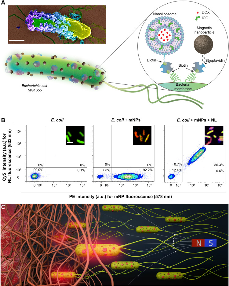
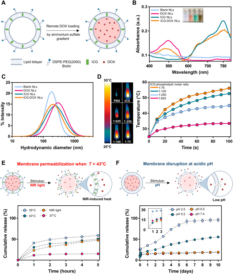
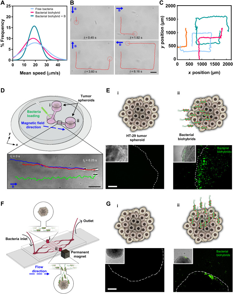
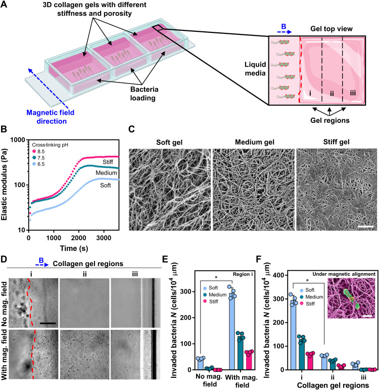
Bacterial biohybrids, composed of self-propelling bacteria carrying micro/nanoscale materials, can deliver their payload to specific regions under magnetic control, enabling additional frontiers in minimally invasive medicine. However, current bacterial biohybrid designs lack high-throughput and facile construction with favorable cargoes, thus underperforming in terms of propulsion, payload efficiency, tissue penetration, and spatiotemporal operation. Here, we report magnetically controlled bacterial biohybrids for targeted localization and multistimuli-responsive drug release in three-dimensional (3D) biological matrices. Magnetic nanoparticles and nanoliposomes loaded with photothermal agents and chemotherapeutic molecules were integrated onto Escherichia coli with ~90% efficiency. Bacterial biohybrids, outperforming previously reported E. coli-based microrobots, retained their original motility and were able to navigate through biological matrices and colonize tumor spheroids under magnetic fields for on-demand release of the drug molecules by near-infrared stimulus. Our work thus provides a multifunctional microrobotic platform for guided locomotion in 3D biological networks and stimuli-responsive delivery of therapeutics for diverse medical applications.


Figures

Fig. 1.
Fig. 1.. Bacterial biohybrids carrying mNPs and NLs.
(A) Schematic illustration of the bacterial biohybrid microrobots, conjugated with NLs and mNPs. NLs are loaded with DOX and ICG, and both NLs and mNPs are conjugated to bacteria via biotin-streptavidin interactions. Inset shows an SEM image of an example bacterial biohybrid carrying mNPs and NLs. Image was pseudocolored. Scale bar, 500 nm. (B) Flow cytometry density plots of (i) free bacteria expressing GFP, (ii) bacterial biohybrids carrying mNPs tagged with red fluorescence, and (iii) bacterial biohybrids carrying mNPs tagged with red fluorescence and NLs tagged with Cy5, showing successful conjugations quantitatively. a.u., arbitrary units. Scale bar, 2 μm. (C) Conceptual schematics depicting bacterial biohybrid microrobots magnetically guided through porous microenvironments toward target tissues, such as a tumor. Bacterial biohybrids can release their payload upon NIR irradiation, enabling stimuli-responsive cargo release in 3D biological matrices. N, north; S, south.
Fig. 2.
Fig. 2.. Synthesis and characterization of DOX- and ICG-loaded NLs.
(A) Schematic of the NL synthesis. ICG is entrapped within the phospholipid bilayer of a NL, and DOX is remotely loaded via the ammonium sulfate gradient method to the inner aqueous core. (B) The absorption spectrum of blank NLs, DOX NLs, ICG NLs, and ICG-DOX NLs. The inset shows the NL solutions placed in PMMA cuvettes. (C) Size distribution of NLs measured by DLS. (D) Infrared thermal images and temperature profiles of various ICG NLs under NIR irradiation (~0.6 W/cm2, 100 s). Different ICG NLs were prepared by changing ICG:phospholipid molar ratio. (E) The NIR-driven cumulative drug release profile of ICG-DOX NLs over 5 hours at 37°, 43°, and 55°C and after NIR irradiation (~0.6 W/cm2, 5 min). At elevated temperatures (>43°C), lipid membranes start to disintegrate and become permeable, inducing DOX release. (F) The pH-driven cumulative drug release profile of DOX NLs over 10 days at pH 2.5, 5.5, 6.5, and 7.4. Low pH triggers membrane disruption and the release of DOX molecules. The inset shows cumulative drug release profile within the first 3 hours of the experiment.
Fig. 3.
Fig. 3.. Motility characterization, external magnetic control, and tumor spheroid localization of magnetic bacterial biohybrids.
(A) 2D swimming velocity analyses of free bacteria and bacterial biohybrids with or without applied external magnetic field. (B) Magnetic control of bacterial biohybrids by changing the applied field (10 mT) direction by 90° turns. B represents the magnetic field vector. Scale bar, 10 μm. (C) 2D swimming trajectories of bacterial biohybrids under external magnetic field control. (D) A magnetic guidance setup with three reservoirs, two of which contain tumor spheroids (i and ii), connected by narrow channels to the loading reservoir. A uniform magnetic field (26 mT) along the x axis was created with a permanent magnet setup. Scale bar, 10 μm. (E) Schematics and microscopy images of the tumor spheroids in reservoirs i and ii. Scale bar, 100 μm. (F) A microfluidic system with branched channels and two reservoirs (i and ii) with one tumor spheroid in each. A small permanent magnet was placed next to the reservoir ii to generate a magnetic field gradient. (G) Schematics and microscopy images of the tumor spheroids in reservoirs i and ii. Scale bar, 100 μm.
Fig. 4.
Fig. 4.. Invasion of collagen gels by magnetically aligned bacterial biohybrids.
(A) Schematic of the experimental setup demonstrating 3D collagen gels with different stiffnesses cross-linked on one side of a tissue culture plate. A permanent magnet setup generating a uniform magnetic field (26 mT), directing bacterial biohybrids toward the collagen gel continuously. (B) Dynamics of collagen gel cross-linking at 37°C. (C) SEM images of 3D collagen gels of varying cross-linking pH. Scale bar, 1 μm. (D) Bright-field optical microscopy images of collagen gel regions after overnight incubation with bacterial biohybrids, with and without magnetic field. Gels were divided into three regions: i, ii, and iii. The red dashed line represents liquid-gel interphase. Gels represented in images were prepared at pH 7.5. Scale bar, 50 μm. (E) Quantification of bacterial biohybrids within three different collagen gels after overnight incubation with or without magnetic field (Student’s t test, P < 0.05). Error bars represent the SD of the mean. (F) Quantification of bacterial biohybrids within three different collagen gels after overnight incubation under uniform magnetic field (Student’s t test, P < 0.05). Error bars represent the SD of the mean. Inset shows a pseudocolored SEM image of a bacterial biohybrid embedded in collagen gel. Scale bar, 1 μm.
Fig. 5.
Fig. 5.. NIR-driven drug release from bacterial biohybrids and drug uptake by tumor spheroids.
(A) Bacterial biohybrids carrying stimuli-responsive ICG-DOX NLs are localized on tumor spheroids and release their payload upon NIR stimulus. NLs convert the light energy into heat, which then prompts phospholipid disintegration and ultimately triggers the release of DOX molecules. (B) DOX delivery to HT-29 tumor spheroids was achieved by coculturing the spheroids with bacterial biohybrids and irradiating the coculture setup with NIR light (~0.6 W/cm2, 10 min). Fluorescence microscopy images show DOX signal from the spheroid 24 hours after NIR irradiation. Scale bar, 100 μm. (C) 3D view of confocal microscopy image shows DOX uptake within the cells of the tumor spheroid 24 hours after NIR irradiation. Scale bar, 75 μm. (D) Fluorescence intensities of free bacteria, bacterial biohybrids carrying DOX NLs, and bacterial biohybrids carrying ICG-DOX NLs, with or without NIR irradiation (Student’s t test, P < 0.05). Error bars represent the SD of the mean. (E) Live/dead viability staining of HT-29 tumor spheroids with bacterial biohybrids carrying ICG-DOX NLs, with and without NIR irradiation. Green and red colors indicate live and dead cells, respectively. Insets show bright-field images of the spheroids. w, with; wo, without. Scale bars, 100 μm.

References

    1. M. Sitti, Mobile Microrobotics (MIT Press, 2017).
    1. Erkoc P., Yasa I. C., Ceylan H., Yasa O., Alapan Y., Sitti M., Mobile microrobots for active therapeutic delivery. Adv. Ther. 2, 1800064 (2019).
    1. Alapan Y., Bozuyuk U., Erkoc P., Karacakol A. C., Sitti M., Multifunctional surface microrollers for targeted cargo delivery in physiological blood flow. Sci. Robot. 5, eaba5726 (2020). - PubMed
    1. Alapan Y., Yigit B., Beker O., Demirörs A. F., Sitti M., Shape-encoded dynamic assembly of mobile micromachines. Nat. Mater. 18, 1244–1251 (2019). - PubMed
    1. Wrede P., Degtyaruk O., Kalva S. K., Deán-Ben X. L., Bozuyuk U., Aghakhani A., Akolpoglu B., Sitti M., Razansky D., Real-time 3D optoacoustic tracking of cell-sized magnetic microrobots circulating in the mouse brain vasculature. Sci. Adv. 8, eabm9132 (2022). - PMC - PubMed