. 2022 Jul 15;377(6603):328-335.
doi: 10.1126/science.abl6324. Epub 2022 Jul 14.

ZAKα-driven ribotoxic stress response activates the human NLRP1 inflammasome

Affiliations

ZAKα-driven ribotoxic stress response activates the human NLRP1 inflammasome

Kim S Robinson et al. Science. .

Abstract

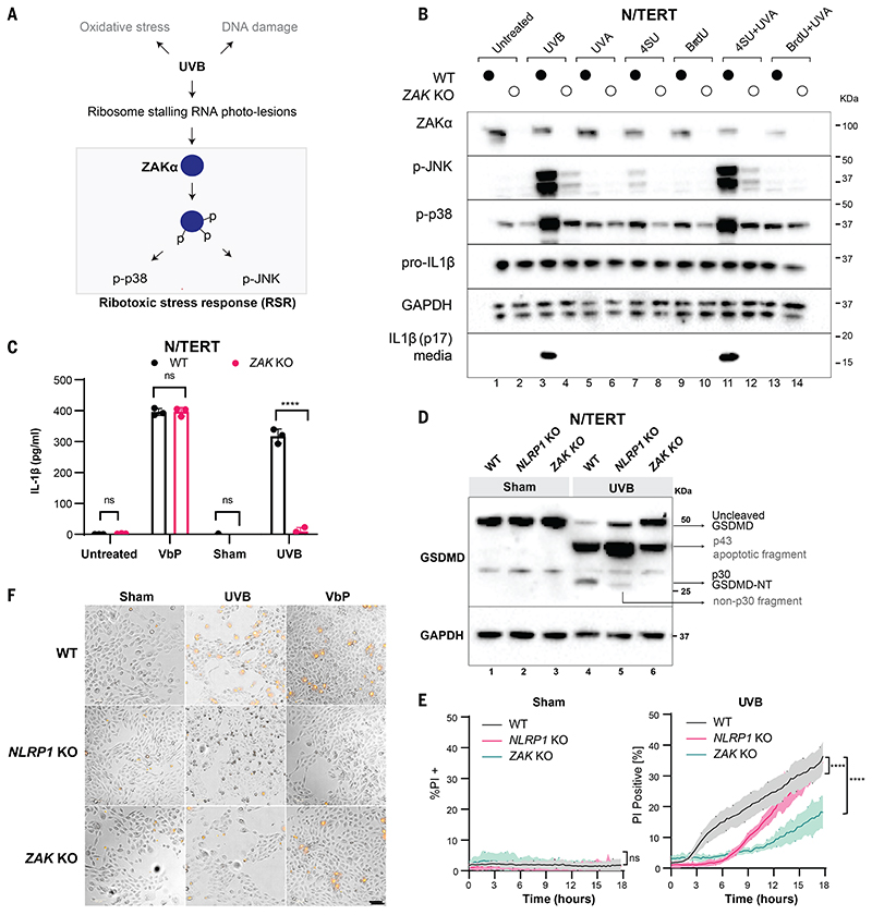
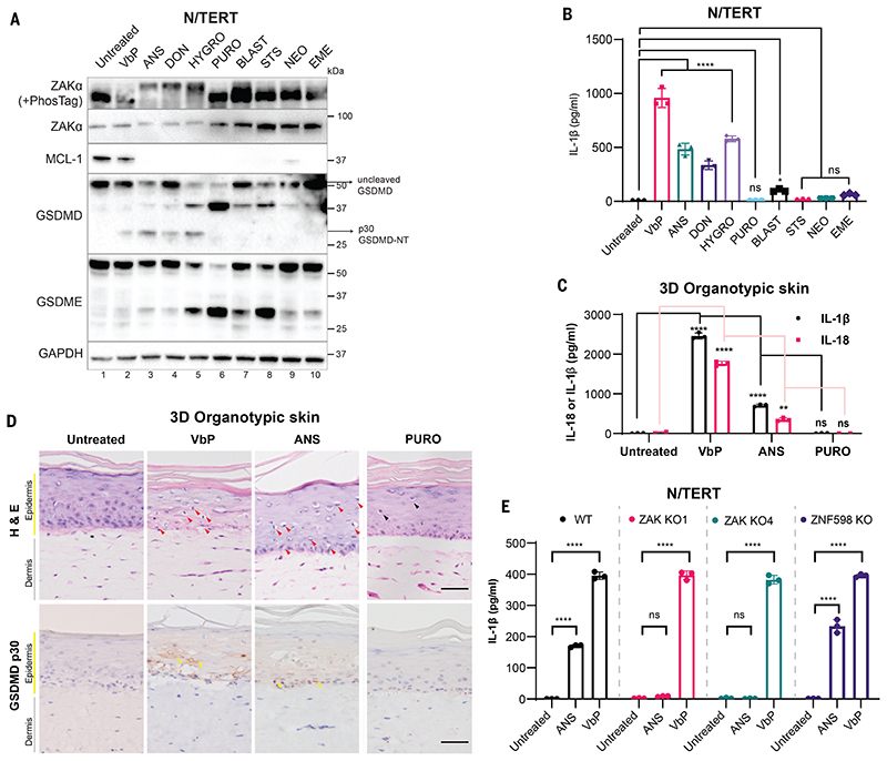
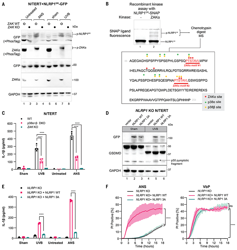
Human NLRP1 (NACHT, LRR, and PYD domain-containing protein 1) is an innate immune sensor predominantly expressed in the skin and airway epithelium. Here, we report that human NLRP1 senses the ultraviolet B (UVB)- and toxin-induced ribotoxic stress response (RSR). Biochemically, RSR leads to the direct hyperphosphorylation of a human-specific disordered linker region of NLRP1 (NLRP1DR) by MAP3K20/ZAKα kinase and its downstream effector, p38. Mutating a single ZAKα phosphorylation site in NLRP1DR abrogates UVB- and ribotoxin-driven pyroptosis in human keratinocytes. Moreover, fusing NLRP1DR to CARD8, which is insensitive to RSR by itself, creates a minimal inflammasome sensor for UVB and ribotoxins. These results provide insight into UVB sensing by human skin keratinocytes, identify several ribotoxins as NLRP1 agonists, and establish inflammasome-driven pyroptosis as an integral component of the RSR.


Conflict of interest statement

Competing interests:

S.L.M. is a scientific advisor for Odyssey therapeutics and NRG therapeutics. J.E.C. is a member of the Board of Directors, 3T Biosciences and Chief Scientific Officer of Parker Institute for Cancer Immunotherapy. F.L.Z., K.S.R., T.G.A., S.Z. are co-inventors of a patent (PCT/SG2022/050086) based on this work.

Figures

Figure 1
Figure 1. ZAKα is required for UVB-triggered NLRP1 inflammasome activation.
(A) Schematic indicating types of cellular damage caused by UVB irradiation. UVB activates ribotoxic stress response (RSR) signaling through ZAKα. (B) Immunoblot of WT N/TERT cells or ZAK KO (sg4) N/TERT cells treated with the indicated combinations of photosensitizer (10 μM) for 4 hours with or without UVA. UVB (100 mJ/cm2) was used as a positive control. *, residual signal after membrane stripping. (C) IL-1β ELISA of WT or ZAK KO (sg4) N/TERT cells after VbP (3 μM) treatment, sham or UVB (100 mJ/cm2) irradiation. Cell culture media were collected 24 hours later. (D) GSDMD immunoblot of WT, NLRP1 KO, ZAK KO (sg4) N/TERT cells treated with UVB (100 mJ/cm2) or sham-irradiated. Cell lysates were harvested 24 hours later. Different GSDMD cleavage fragments are shown by black arrows. Note that the GSDMD antibody used in this experiment recognizes all GSDMD-cleaved products. In NLRP1 KO cells UVB leads to a weak band <30 kDa. (E) Quantification of the percentage of PI-positive WT, NLRP1 KO, ZAK KO (sg4) N/TERT cells after sham or UVB (100 mJ/cm2) irradiation. (F) Representative images of PI inclusion 5 hours post irradiation from 3 independent experiments. Scale bar represents 100 μm. Error bars represent standard errors of the mean (S.E.M.) from three biological replicates, where one replicate refers to an independent seeding and treatment of the cells. The significance values were calculated based on two-way ANOVA followed by Sidak’s test for multiple pairwise comparisons in (C), and two-tailed Kolmogorov-Smirnov test at 95% confidence interval in (E). ns, non-significant, ****P<0.0001.
Figure 2
Figure 2. ZAKα-activating compounds induce NLRP1-driven pyroptosis.
(A) Immunoblot of N/TERT cell lysate after treatment with the indicated drugs. ZAKα phosphorylation was detected after 3 hours of drug treatment, while immunoblots for MCL-1, GSDMD, and GSDME were performed using samples 24 hours post treatment. (B) IL-1β ELISA of N/TERT cell media 24 hours post treatment with the indicated drugs at concentrations specified in A. Note that a smaller volume of the media and a higher number of cells were used in this experiment, accounting for the overall higher concentration of IL-1β. (C) IL-1β and IL-18 ELISA of growth media collected from 3D organotypic skin cultures treated with the indicated drugs. (D) H&E and cleaved GSDMD-NT (p30 specific) immunostaining of 3D organotypic skin cultures treated with the indicated drugs in D. Scale bar represents 100 μm. Red arrows indicate keratinocytes with diminished eosin but dense hematoxylin staining that were abundant in VbP- and ANS-treated cultures. Black arrows indicate putatively apoptotic cells with low eosin and hematoxylin staining that were abundant in PURO-treated samples. Yellow arrows indicate membranous GSDMD p30 staining. Images represent one of 3 independent organotypic skin cultures. (E) IL-1β ELISA of culture media from N/TERT cells of the indicated genotypes after 24 hours of drug treatment. VbP was used at 3 μM and ANS at 1 μM. Error bars represent standard errors of the mean (S.E.M.) from three biological replicate experiments, where one replicate refers to an independent seeding and treatment of the cells. The significance values were calculated based on two-way ANOVA followed by Dunnett’s test for multiple pairwise comparisons in (B) and (C), and Sidak’s test in (E). ns, non-significant, *P<0.05, **P<0.01, ****P<0.0001.
Figure 3
Figure 3. A disordered linker region of human NLRP1 selectively mediates ZAKα-dependent activation.
(A) IL-1β ELISA from NLRP1 KO N/TERT cells reconstituted with the indicated NLRP1 variants and CARD8 treated with ANS (1 μM) and VbP (3 μM). Note that this experiment was performed independently using higher cell numbers. (B) Comparison between the domain structures of human NLRP1 and rodent NLRP1a-c. The predicted disorder score was calculated for a.a. 1-300 of human NLRP1. (C) IL-1β ELISA from NLRP1-KO N/TERT cells reconstituted with GFP-full-length NLRP1 or NLRP1 lacking PYD+DR (a.a.1-254). Cells were treated with the indicated drugs, or sham- or UVB-irradiated and harvested 24 hours post-treatment. (D) Comparison of the domain arrangements of human NLRP1 and CARD8 and the engineered hybrid sensor referred to as NLRP1DR-CARD8ZC. (E) IL-1β ELISA from NLRP1-KO N/TERT cells transduced with CARD8 or NLRP1DR-CARD8ZC and treated with 1 μM ANS or 3 μM VbP for 24 hours. (F) GSDMD and IL-1β immunoblot from the cells in E, along with WT or ZAK KO N/TERT cells irradiated with UVB. The GSDMD antibody recognizes both full-length and cleaved forms, including p43 and p30. The IL-1β immunoblot was performed with samples that combined lysate and 10 times concentrated media. The error bars represent standard errors of the mean (S.E.M.) from three biological replicates, where one replicate refers to an independent seeding and treatment of the cells. The significance values in (A), (C) and (E) were calculated based on two-way ANOVA followed by Sidak’s test for multiple pairwise comparisons. Significance values were indicated as: ns, non-significant, **P<0.01, *** P<0.001, ****P<0.0001.
Figure 4
Figure 4. Hyperphosphorylation of NLRP1DR by ZAKα and p38 activates NLRP1.
(A) Immunoblot following SDS-PAGE or PhosTag SDS-PAGE of wild-type or ZAK-KO N/TERT cells expressing NLRP1DR-GFP. Cells were harvested 2 hours post ANS treatment or UVB irradiation. (B) Recombinant SNAP-tagged NLRP1DR was incubated with recombinant ZAKα in a standard kinase reaction for 30 min. NLRP1DR phosphorylation was visualized with SNAP ligand fluorescence (TMR) on a PhosTag-containing SDS-PAGE gel. (C) IL-1β ELISA from WT, MAPK14 MAPK11 DKO (denoted as p38α+β DKO) and ZAK KO N/TERT cells 24 hours after UVB irradiation or ANS treatment. (D) GFP and GSDMD immunoblot of NLRP1-KO N/TERT cells expressing full-length WT NLRP1 or full-length NLRP1 T178A, S179A, T180A (3A) mutant after UVB irradiation. All constructs were fused with GFP at the N- terminus. GSDMD p30 is marked with a black arrow. Cells were harvested 24 hours post UVB treatment. (E) IL-1β ELISA from NLRP1-KO N/TERT cells expressing full length WT NLRP1 or full-length NLRP1 T178A, S179A, T180A (3A) mutant 24 hours after UVB irradiation or ANS treatment. (F) Quantification of the percentage of PI+ NLRP1-KO N/TERT cells expressing full-length WT NLRP1 or full-length NLRP1 T178A, S179A, T180A (3A) mutants in the presence of ANS or VbP. Images were acquired at 15-min intervals for 18 hours. Error bars represent standard errors of the mean (S.E.M.) from three biological replicates, where one replicate refers to an independent seeding and treatment of the cells. The significance values were calculated based on two-way ANOVA followed by Sidak’s test for multiple pairwise comparisons in (C) and (E), and two-tailed Kolmogorov-Smirnov test at 95% confidence interval in (F). ns, non-significant, ****P<0.0001.

References

    1. Rathinam VAK, Fitzgerald KA. Inflammasome Complexes: Emerging Mechanisms and Effector Functions. Cell. 2016;165:792–800. - PMC - PubMed
    1. Broz P, Dixit VM. Inflammasomes: mechanism of assembly, regulation and signalling. Nat Rev Immunol. 2016;16:407–420. - PubMed
    1. Man SM, Kanneganti T-D. Regulation of inflammasome activation. Immunol Rev. 2015;265:6–21. - PMC - PubMed
    1. Taabazuing CY, Griswold AR, Bachovchin DA. The NLRP1 and CARD8 inflammasomes. Immunol Rev. 2020;297:13–25. - PMC - PubMed
    1. Bachovchin DA. NLRP1: a jack of all trades, or a master of one? Mol Cell. 2021;81:423–425. - PubMed

MeSH terms