. 2022 Aug 12;377(6607):735-742.
doi: 10.1126/science.abq2679. Epub 2022 Jul 12.

ACE2-binding exposes the SARS-CoV-2 fusion peptide to broadly neutralizing coronavirus antibodies

Affiliations

ACE2-binding exposes the SARS-CoV-2 fusion peptide to broadly neutralizing coronavirus antibodies

Jun Siong Low et al. Science. .

Abstract

The coronavirus spike glycoprotein attaches to host receptors and mediates viral fusion. Using a broad screening approach, we isolated seven monoclonal antibodies (mAbs) that bind to all human-infecting coronavirus spike proteins from severe acute respiratory syndrome coronavirus 2 (SARS-CoV-2) immune donors. These mAbs recognize the fusion peptide and acquire affinity and breadth through somatic mutations. Despite targeting a conserved motif, only some mAbs show broad neutralizing activity in vitro against alpha- and betacoronaviruses, including animal coronaviruses WIV-1 and PDF-2180. Two selected mAbs also neutralize Omicron BA.1 and BA.2 authentic viruses and reduce viral burden and pathology in vivo. Structural and functional analyses showed that the fusion peptide-specific mAbs bound with different modalities to a cryptic epitope hidden in prefusion stabilized spike, which became exposed upon binding of angiotensin-converting enzyme 2 (ACE2) or ACE2-mimicking mAbs.


Figures
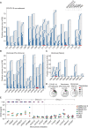
Fig. 1.
Fig. 1.. Rare broadly reactive memory B cells are elicited upon natural infection or vaccination.
(A to D) Total PBMCs from COVID-19 convalescents (C) (A), vaccinees with prior infection (pre-immune) (VP) (B), and vaccinees without prior infection (naïve) (VN) (C) (table S5) were plated in replicate 96 wells (104 cells/well) and stimulated with TLR agonist R848 (2.5 μg/ml) in the presence of IL-2 (1,000 U/ml). Twelve days later, the supernatant of each culture was screened, in parallel, for the specificities of the secreted IgG antibodies to commercially available hCoV S proteins from SARS-CoV-2, SARS-CoV, OC43, HKU1, NL63 and 229E by ELISA. The skyline plot provides a detailed view of the specificities of each culture well (represented in rows) to the respective antigens (in subcolumn) from each donor (in column). The order of the antigens is indicated in the legend and uncoated plates (PBS) were used as control. If OD 405nm value exceeds the cut-off value determined by average OD 405nm of PBS wells + 4*standard deviation, the culture was considered reactive to the antigen and indicated with colored cells. Only cultures exhibiting reactivity to at least one antigen are shown. Red cells highlight the six cultures exhibiting reactivity to all alpha and beta hCoV S proteins tested. (D) Cross-reactivity patterns of SARS-CoV-2 S positive cultures from (A to C) shown as pie charts. The total number of SARS-CoV-2 S-positive cultures from each cohort is indicated at the center of the pie. (E) Using a two-step screening strategy as depicted in fig. S1, 16 mAbs that cross-reacted with multiple hCoV S proteins were isolated from 10 donors (C series from convalescent donors, VP and VN series from pre-immune and naïve vaccinees). Shown are EC50 values to the respective commercially available hCoV S protein, measured by ELISA. mAbs are grouped based on the reactivity patterns. Shown is one representative experiment out of at least 2 performed. Group 2 mAbs C21G12, C21D10, and C21E3 were described in a separate study ( 15 ) (designated P34G12, P34D10, and P34E3, respectively).
Fig. 2.
Fig. 2.. Broadly reactive fusion peptide-targeted mAbs acquire affinity and breadth through somatic mutations.
(A) Binding profiles of the seven Group 3 mAbs. Epitope mapping was performed using commercially available SARS-CoV-2 protein domains (S1, RBD, S2), 15-mer overlapping synthetic peptides with the indicated sequences, prefusion HexaPro (F817P) S, prefusion PentaPro (F817) S, prefusion PentaPro (F817) S2 and postfusion S2 coated onto plastic and assessed by ELISA (log10 EC50 shown). The mAbs all recognize the fusion peptide sequence highlighted in blue. (B) Binding evaluation of Group3 mAbs and their respective germline reverted unmutated common ancestors (UCAs) to different hCoV S proteins. C13B8 and C13A7 are sister clones and thus share a single UCA. One representative experiment out of at least two experiments is shown. (C) Half-maximum inhibitory concentrations (IC50s) of Group 3 mAbs against SARS-CoV-2, SARS-CoV, MERS-CoV, NL63 and 229E pseudoviruses in the indicated target cell lines. For each hCoV pseudotyped assay, all mAbs were compared in parallel. One representative experiment out of at least two experiments is shown. (D) Group 3 mAbs were assessed for their ability to inhibit the fusion of SARS-CoV-2 S-expressing (A549-S) and ACE2-TMPRSS2-expressing (A549-ACE2-TMPRSS2) A549 cells. Inhibition of fusion values are normalized to the percentage of fusion without mAb (100%) and to that of fusion of non-transfected cells (0%). One representative experiment out of two is shown. (E) The prophylactic efficacy of VN01H1 (50 mg/kg), C77G12 (25 mg/kg and 50 mg/kg) and negative control anti-malaria mAb MGH2 (50 mg/kg) were tested in hamsters challenged with the SARS-CoV-2 P.1 (Gamma) variant of concern. Viral RNA loads (top), replicating virus titers (middle), and histopathological scores (bottom) are shown. ns P > 0.05, *P ≤ 0.05, **P ≤ 0.01, ***P ≤ 0.001, Mann-Whitney test corrected using Bonferroni multiple comparison.
Fig. 3.
Fig. 3.. Fusion peptide antibodies target a cryptic epitope.
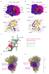
(A and B) (Top) Surface representation of the crystal structures of the C77G12 (A) and VN01H1 (B) Fabs in complex with SARS-CoV-2 fusion peptide epitope. (Bottom) Ribbon representation of the corresponding structures highlighting the interactions of Fab heavy and light chain CDRs with the fusion peptide (selected regions are shown for clarity). (C), Ribbon representation of the fusion peptides in the Fab-bound complexes superimposed with the fusion peptide in prefusion SARS-CoV-2 S (PDB 6VXX). (D and E) Superimposition of the C77G12-bound (D) or VN01H1-bound (E) fusion peptide structures to prefusion SARS-CoV-2 S uncovering the cryptic nature of the epitope. The Fabs are shown as surfaces whereas S is rendered as ribbons. Each SARS-CoV-2 S protomer is colored distinctly (light blue, pink, and gold) and Fab heavy and light chains are colored purple and magenta, respectively.
Fig. 4.
Fig. 4.. The SARS-CoV-2 fusion peptide is unmasked following ACE2 receptor engagement.
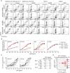
(A) Binding of fusion peptide-specific mAbs VN01H1 and C77G12 (8 μg/ml), as well as RBD-specific C94 and stem helix-specific C21E3 mAbs (8 μg/ml) on HEK293T cells transiently co-transfected with plasmid encoding ZsGreen and plasmids encoding full length SARS-CoV-2 S Wuhan-Hu-1 (GenBank: NC_045512), SARS-CoV-2 S-2P (K986P, V987P), NL63 S (GenBank: APF29071.1) or 229E S (GenBank: APT69883.1), in the presence or absence of receptor ACE2 or APN, as measured by flow cytometry. (B) Titrating doses of fluorescently labeled mAbs were co-incubated with HEK293T cells expressing full-length SARS-CoV-2 S for 2 hours at room temperature in the presence or absence of recombinant ACE2-mFc (27 μg/ml), ACE2-mimicking mAb S2E12 (20 μg/ml) or S2M11 (20 μg/ml), the latter mAb locks the S trimer in a closed conformation. The line in the scatter plot is a reference for the maximum gMFI of staining with control anti-RBD C94 mAb. One representative experiment out of two is shown. (C) VN01H1 and C77G12 in full-length IgG or scFv formats were tested for their neutralization of authentic SARS-CoV-2 Washington-1, Omicron BA.1 and BA.2 variants. IC50 values are displayed as a table. (D) Synergy experiment was performed against SARS-CoV-2 (Wuhan-Hu-1) pseudovirus (0.1 MOI) on VeroE6-TMPRSS2 at indicated concentrations of S2E12 and C77G12 mAbs alone or in combination, in a checkerboard manner. Analysis of mAb synergy was carried out using MacSynergy II ( 70 ). Synergy plots adjusted with Bonferroni correction at 95% confidence were used for reporting. A synergy score between 50 and 100 was defined as moderate.

References

    1. Vlasova A. N., Diaz A., Damtie D., Xiu L., Toh T.-H., Lee J. S.-Y., Saif L. J., Gray G. C., Novel canine coronavirus isolated from a hospitalized patient with pneumonia in East Malaysia. Clin. Infect. Dis. 74, 446–454 (2022). 10.1093/cid/ciab456 - DOI - PMC - PubMed
    1. Lednicky J. A., Tagliamonte M. S., White S. K., Elbadry M. A., Alam M. M., Stephenson C. J., Bonny T. S., Loeb J. C., Telisma T., Chavannes S., Ostrov D. A., Mavian C., Beau De Rochars V. M., Salemi M., Morris J. G. Jr., Independent infections of porcine deltacoronavirus among Haitian children. Nature 600, 133–137 (2021). 10.1038/s41586-021-04111-z - DOI - PMC - PubMed
    1. Tortorici M. A., Walls A. C., Joshi A., Park Y.-J., Eguia R. T., Miranda M. C., Kepl E., Dosey A., Stevens-Ayers T., Boeckh M. J., Telenti A., Lanzavecchia A., King N. P., Corti D., Bloom J. D., Veesler D., Structure, receptor recognition, and antigenicity of the human coronavirus CCoV-HuPn-2018 spike glycoprotein. Cell 185, 2279–2291.e17 (2022). 10.1016/j.cell.2022.05.019 - DOI - PMC - PubMed
    1. Walls A. C., Park Y.-J., Tortorici M. A., Wall A., McGuire A. T., Veesler D., Structure, function, and antigenicity of the SARS-CoV-2 spike glycoprotein. Cell 181, 281–292.e6 (2020). 10.1016/j.cell.2020.02.058 - DOI - PMC - PubMed
    1. Wrapp D., Wang N., Corbett K. S., Goldsmith J. A., Hsieh C.-L., Abiona O., Graham B. S., McLellan J. S., Cryo-EM structure of the 2019-nCoV spike in the prefusion conformation. Science 367, 1260–1263 (2020). 10.1126/science.abb2507 - DOI - PMC - PubMed

MeSH terms